Lumbar vertebrae
Five large weight-bearing vertebrae of the lower back. Spondylolysis, spondylolisthesis and lumbar spinal stenosis are their key clinical problems.
Core anatomy
- Location
Lower back, L1–L5, between T12 and the sacrum.
- Classification
Irregular bones.
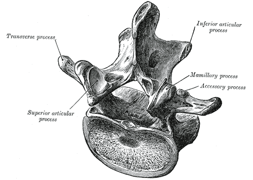
- Key features & landmarks
- Large, kidney-shaped bodies (the heaviest load)
- Short, thick, hatchet-shaped spinous processes (easy to palpate; interspaces widen in flexion for lumbar puncture)
- Articular facets oriented roughly sagittally, allowing flexion and extension but little rotation
- Mammillary and accessory processes (muscle attachments)
- Pars interarticularis: the bone between the superior and inferior articular processes, the site of stress fractures
- Triangular vertebral foramen containing the conus medullaris (L1) and cauda equina
- Relations
Lumbar nerve roots exit below the pedicle of the same-numbered vertebra (L4 exits through the L4–L5 foramen). The psoas major arises from the sides of the bodies; the abdominal aorta and inferior vena cava lie anteriorly.
Swipe sideways to see the whole diagram →
Swipe sideways to see the whole diagram →

Clinical correlations
Spondylolysis PANCE
- Mechanism / lesion
Stress fracture of the pars interarticularis, most often at L5, from repetitive hyperextension in adolescent athletes (gymnastics, football linemen, diving, dance).
- Classic presentation
Low back pain in an adolescent athlete, worse with extension. Positive single-leg hyperextension (stork) test. On oblique X-ray, a "collar" on the "Scottie dog".
Spondylolisthesis PANCE
- Mechanism / lesion
Forward slip of one vertebra on the one below. Isthmic (from bilateral spondylolysis, typically L5 on S1) in younger patients; degenerative (facet arthropathy, typically L4 on L5) in older adults, especially women.
- Classic presentation
Low back pain, hamstring tightness, radicular symptoms, and a palpable step-off of the spinous processes. Graded by percentage of slip on a lateral X-ray.
Lumbar spinal stenosis PANCE
- Mechanism / lesion
Narrowing of the central canal, lateral recesses or foramina from disc bulging, facet hypertrophy and ligamentum flavum thickening, usually in adults over 50.
- Classic presentation
Neurogenic claudication: buttock and leg pain, numbness or weakness on walking and standing (extension), relieved by sitting or bending forward (the "shopping cart sign": leaning on a cart helps). Pedal pulses are normal (unlike vascular claudication).
High-yield PANCE pearls
- Adolescent athlete + back pain with extension → spondylolysis (L5 pars); Scottie dog collar.
- Spondylolisthesis: step-off; isthmic L5–S1 (young), degenerative L4–L5 (older).
- Spinal stenosis: pain with walking/standing, relief with flexion/sitting; MRI; normal pulses.
- Lumbar roots exit below their same-numbered pedicle.
Imaging anatomy
- X-ray
Lateral view grades spondylolisthesis; oblique views show the pars (the "Scottie dog": its neck is the pars, so a lucent "collar" indicates spondylolysis). Flexion–extension views show instability. X-rays aren't needed for acute nonspecific low back pain without red flags.
- CT
Best to confirm pars fractures and assess healing.
- MRI
First choice for spinal stenosis and radiculopathy (canal and foraminal narrowing, nerve root compression); STIR shows early pars stress reaction.
- Key views & landmarks
- Standing lateral lumbar radiograph (spondylolisthesis)
- MRI lumbar spine (stenosis, radiculopathy with red flags or persistent symptoms)
Physical exam
- Surface landmarks
The iliac crests mark about L4; the L5 spinous process lies just above the sacrum.
- Palpation
Step-off between spinous processes (spondylolisthesis); paraspinal tenderness.
- Strength / function testing
Lumbar range of motion (pain with extension suggests posterior element pathology; with flexion, disc). Lower limb neurologic exam and pulses (neurogenic vs vascular claudication).
Single-leg hyperextension (stork) test
- Technique
The patient stands on one leg and extends the lumbar spine (± rotation toward the stance side).
- Positive test
Reproduction of low back pain on the stance side.
- Significance
Suggests spondylolysis (pars stress injury), though it isn't specific.
Self-quiz
A 15-year-old gymnast has 2 months of low back pain that worsens with back walkovers and standing on one leg while arching her back. What is the most likely diagnosis?
Correct answer: B
Spondylolysis (a pars interarticularis stress fracture, usually L5) is typical in adolescents doing repetitive hyperextension. The stork test reproduces the pain.
Show answer
B
Spondylolysis (a pars interarticularis stress fracture, usually L5) is typical in adolescents doing repetitive hyperextension. The stork test reproduces the pain.
A 72-year-old man has buttock and leg pain after walking two blocks that improves when he sits or leans forward on a shopping cart. Pedal pulses are normal. What is the most likely diagnosis?
Correct answer: B
Neurogenic claudication relieved by flexion (sitting, leaning forward) with normal pulses is classic for lumbar spinal stenosis. Vascular claudication is relieved by standing still and has reduced pulses.
Show answer
B
Neurogenic claudication relieved by flexion (sitting, leaning forward) with normal pulses is classic for lumbar spinal stenosis. Vascular claudication is relieved by standing still and has reduced pulses.
On an oblique lumbar radiograph, a lucent defect across the "neck of the Scottie dog" represents a defect in which structure?
Correct answer: C
The Scottie dog's neck is the pars interarticularis; a lucent "collar" indicates spondylolysis.
Show answer
C
The Scottie dog's neck is the pars interarticularis; a lucent "collar" indicates spondylolysis.
A 68-year-old woman has back pain and neurogenic claudication. Lateral radiographs show a degenerative spondylolisthesis. At which level is this most typical?
Correct answer: C
Degenerative spondylolisthesis (from facet arthropathy) typically occurs at L4 on L5 in older adults, especially women. Isthmic spondylolisthesis (from bilateral spondylolysis) typically occurs at L5 on S1 in younger patients.
Show answer
C
Degenerative spondylolisthesis (from facet arthropathy) typically occurs at L4 on L5 in older adults, especially women. Isthmic spondylolisthesis (from bilateral spondylolysis) typically occurs at L5 on S1 in younger patients.
The roughly sagittal orientation of the lumbar facet joints allows which movements?
Correct answer: A
Lumbar facets are oriented roughly sagittally, allowing flexion and extension but little rotation. Thoracic facets lie roughly in the coronal plane, favoring rotation (limited by the ribs).
Show answer
A
Lumbar facets are oriented roughly sagittally, allowing flexion and extension but little rotation. Thoracic facets lie roughly in the coronal plane, favoring rotation (limited by the ribs).