Ductus deferens and seminal glands
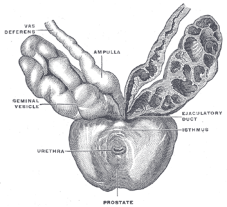
Thick-walled muscular duct carrying sperm from the epididymis to the ejaculatory duct, joined behind the bladder by the seminal gland. It is cut in vasectomy and absent in men with cystic fibrosis.
Core anatomy
- Location
From the tail of the epididymis up the back of the testis and epididymis in the spermatic cord, through the inguinal canal, then across the lateral pelvic wall and behind the urinary bladder to the base of the prostate. About 45 cm long.
- Parts
- Scrotal part: easily palpable as a firm cord (the site of vasectomy)
- Inguinal part: within the spermatic cord in the inguinal canal
- Pelvic part: at the deep inguinal ring it leaves the cord, hooks lateral to the inferior epigastric artery, crosses the external iliac vessels, and passes over the ureter near the bladder
- Ampulla: dilated end behind the bladder, medial to the seminal gland
- Seminal glands (vesicles): coiled glands lateral to the ampullae, behind the bladder; secrete an alkaline, fructose-rich fluid forming most of the semen volume
- Ejaculatory ducts: formed by the union of the ductus and the seminal gland duct; pass through the prostate to open on the seminal colliculus of the prostatic urethra
- Relations
The ureter passes under the ductus deferens near the posterolateral bladder (the male "water under the bridge"). The ampullae and seminal glands lie against the anterior wall of the rectum.
- Arterial supply
Artery to the ductus deferens (usually from the superior or inferior vesical artery, anterior division of the internal iliac artery); seminal glands from the inferior vesical and middle rectal arteries.
- Venous drainage
Pampiniform plexus (scrotal part) and the vesical/prostatic venous plexuses.
- Lymphatic drainage
External and internal iliac nodes.
- Innervation
Sympathetic fibers (about L1–L2) via the hypogastric plexuses cause the peristaltic contraction of the ductus and seminal glands during emission. See pelvic autonomic nerves.
Mentioned in
Generated automatically from links in other entries.

Clinical correlations
Vasectomy PANCE
- Mechanism / lesion
Division or occlusion of the scrotal ductus deferens bilaterally.
- Classic presentation
Ejaculate volume is almost unchanged (most comes from the seminal glands and prostate). Sperm remain downstream for weeks, so contraception continues until a post-vasectomy semen analysis confirms azoospermia (the AUA allows testing from 8 weeks after vasectomy).
Congenital bilateral absence of the vas deferens (CBAVD) PANCE
- Mechanism / lesion
Associated with CFTR mutations — present in almost all men with cystic fibrosis.
- Classic presentation
Obstructive azoospermia with low ejaculate volume and acidic semen (no seminal gland fluid); the ductus cannot be felt in the scrotum. Offer CFTR testing to the patient and partner.
Injury during inguinal hernia repair
- Mechanism / lesion
The ductus runs through the inguinal canal in the spermatic cord.
- Classic presentation
Obstruction (unilateral obstructive azoospermia contributes to infertility).
High-yield PANCE pearls
- Male infertility with absent vas deferens on exam → cystic fibrosis/CFTR testing.
- After vasectomy, use contraception until semen analysis shows azoospermia.
- Emission = sympathetic (ductus, seminal glands, prostate contract).
- Seminal glands supply most of the semen volume (fructose).
Imaging anatomy
- MRI
Shows seminal gland invasion in prostate cancer staging.
- Ultrasound
Transrectal ultrasound shows dilated seminal glands or ejaculatory duct cysts in ejaculatory duct obstruction, and absent or hypoplastic seminal glands in CBAVD. Scrotal ultrasound shows the epididymis and scrotal ductus.
Physical exam
- Surface landmarks
The scrotal ductus is palpable in the posterior part of the spermatic cord between the testis and the superficial inguinal ring.
- Palpation
Roll the spermatic cord between the thumb and fingers: the ductus is a firm, smooth "whipcord". Its absence in an infertile man suggests CBAVD. Normal seminal glands are not palpable on DRE.
Self-quiz
A 30-year-old man is evaluated for infertility. Semen analysis shows azoospermia with low ejaculate volume and acidic pH. On exam, neither ductus deferens can be palpated. Which test is most appropriate?
Correct answer: B
Congenital bilateral absence of the vas deferens causes obstructive azoospermia with low-volume, acidic ejaculate (missing seminal gland fluid) and is strongly associated with CFTR mutations. Testing the patient (and his partner before assisted reproduction) is indicated.
Show answer
B
Congenital bilateral absence of the vas deferens causes obstructive azoospermia with low-volume, acidic ejaculate (missing seminal gland fluid) and is strongly associated with CFTR mutations. Testing the patient (and his partner before assisted reproduction) is indicated.
Near the posterolateral bladder, the ductus deferens crosses which structure?
Correct answer: B
The ductus deferens passes over (superior/anterior to) the ureter — the ureter is "under the bridge", as with the uterine artery in females.
Show answer
B
The ductus deferens passes over (superior/anterior to) the ureter — the ureter is "under the bridge", as with the uterine artery in females.
After vasectomy, why is ejaculate volume almost unchanged?
Correct answer: B
The seminal glands (fructose-rich fluid) and the prostate supply most of the semen volume, so dividing the scrotal ductus barely changes volume. Contraception continues until a semen analysis confirms azoospermia.
Show answer
B
The seminal glands (fructose-rich fluid) and the prostate supply most of the semen volume, so dividing the scrotal ductus barely changes volume. Contraception continues until a semen analysis confirms azoospermia.