Dural venous sinuses
Valveless venous channels between the layers of the dura that drain the brain into the internal jugular veins. Thrombosis of the sagittal or cavernous sinus is an important, easily missed cause of headache and cranial nerve palsies.
Core anatomy
- Formation
Endothelium-lined spaces between the periosteal and meningeal layers of the dura (see cranial meninges and intracranial hemorrhage); they receive the cerebral, cerebellar, diploic and emissary veins and CSF from the arachnoid granulations.
- Course
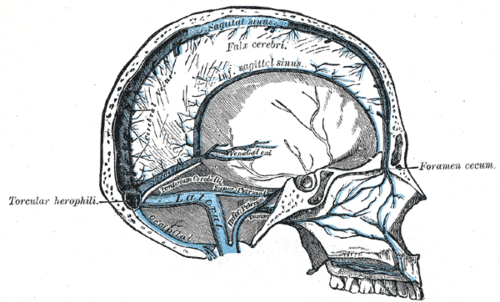
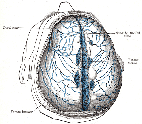
Superior sagittal sinus (along the attached border of the falx; receives CSF via arachnoid granulations) and straight sinus (formed by the inferior sagittal sinus and the great cerebral vein of Galen) meet at the confluence of sinuses (near the internal occipital protuberance) → transverse sinuses → sigmoid sinuses (grooving the mastoid part of the temporal bone) → internal jugular vein at the jugular foramen. The cavernous sinuses on each side of the sella drain via the superior and inferior petrosal sinuses.
- Tributaries
- Superior cerebral veins (bridging veins) → superior sagittal sinus
- Great cerebral vein (of Galen) → straight sinus
- Superior and inferior ophthalmic veins → cavernous sinus (connecting it with the facial veins)
- Emissary veins (scalp ↔ sinuses) and diploic veins
- Drains into
Internal jugular vein (via the sigmoid sinus and inferior petrosal sinus).
- Relations
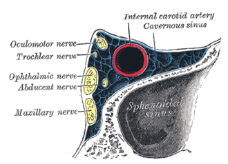
Cavernous sinus contents: internal carotid artery and CN VI run through the sinus; CN III, IV, V1 and V2 run in its lateral wall (see oculomotor nerve (CN III), trochlear nerve (CN IV), trigeminal nerve (CN V), abducens nerve (CN VI)); the pituitary gland lies medially.
- Notes
The valveless facial veins (angular → ophthalmic) connect the "danger triangle" of the face (nose and upper lip) with the cavernous sinus; scalp emissary veins connect scalp infections with the intracranial sinuses (see scalp).
Mentioned in
- Abducens nerve (CN VI)
- Cerebral cortex and lobes
- Oculomotor nerve (CN III)
- Orbit
- Pituitary gland
- Trigeminal nerve (CN V)
- Trochlear nerve (CN IV)
- Ventricular system and CSF
Generated automatically from links in other entries.
Swipe sideways to see the whole diagram →



Clinical correlations
Cerebral venous sinus thrombosis PANCE
- Mechanism / lesion
Prothrombotic states — oral contraceptives, pregnancy and postpartum, thrombophilias, cancer, dehydration, infection (mastoiditis → sigmoid/transverse sinus).
- Classic presentation
Headache (often progressive, worse lying down), papilledema, seizures, focal deficits, altered consciousness; venous (often hemorrhagic) infarcts not matching arterial territories. Diagnose with MR or CT venography (empty delta sign on contrast CT). Treat with anticoagulation (heparin) even if there is hemorrhage.
Cavernous sinus thrombosis PANCE
- Mechanism / lesion
Septic thrombosis spreading from the face (danger triangle), sphenoid/ethmoid sinusitis, orbit or teeth; usually S. aureus.
- Classic presentation
Fever, headache, proptosis, chemosis, periorbital edema, ophthalmoplegia (CN VI often first), reduced sensation in V1/V2, and spread to the other eye. IV antibiotics ± anticoagulation; MRI/CT venography.
Carotid–cavernous fistula
- Mechanism / lesion
Abnormal connection between the ICA (or its branches) and the cavernous sinus — trauma or ruptured cavernous aneurysm.
- Classic presentation
Pulsatile exophthalmos, orbital bruit, chemosis, arterialized conjunctival veins, ophthalmoplegia; endovascular repair.
High-yield PANCE pearls
- Headache + papilledema + seizures in a woman on OCPs or postpartum → venous sinus thrombosis → MRV → heparin (even with hemorrhage).
- Facial/sinus infection → proptosis + chemosis + ophthalmoplegia + fever → cavernous sinus thrombosis.
- CN VI is the first/most affected nerve in the cavernous sinus.
- Pulsatile exophthalmos + bruit → carotid–cavernous fistula.
- Sinus drainage: superior sagittal → confluence → transverse → sigmoid → IJV.
Imaging anatomy
- CT
Noncontrast CT may show a hyperdense sinus ("cord sign") or venous infarcts; CT venography shows filling defects (empty delta sign).
- MRI
MRI with MR venography — the test of choice for venous sinus thrombosis; contrast MRI of the orbits and cavernous sinus.
- Key views & landmarks
- MR venography
- CT venography
Physical exam
- Strength / function testing
Fundoscopy (papilledema), eye movements (CN III, IV, VI), corneal reflex and V1/V2 sensation, proptosis and chemosis, facial skin and sinuses for an infective source.
Self-quiz
A 29-year-old woman 2 weeks postpartum has a worsening headache for 5 days, blurred vision and a seizure. Fundi show bilateral papilledema. CT shows a small hemorrhagic infarct in the left parietal lobe. What is the most likely diagnosis?
Correct answer: B
Postpartum hypercoagulability, headache, papilledema, seizures and a hemorrhagic venous infarct suggest cerebral venous sinus thrombosis. Confirm with MR/CT venography; treat with anticoagulation despite the hemorrhage.
Show answer
B
Postpartum hypercoagulability, headache, papilledema, seizures and a hemorrhagic venous infarct suggest cerebral venous sinus thrombosis. Confirm with MR/CT venography; treat with anticoagulation despite the hemorrhage.
A 35-year-old squeezed a boil on the side of his nose. Three days later he has fever, proptosis, chemosis and cannot abduct the right eye. Which structure is involved?
Correct answer: B
Infection from the danger triangle spreads through the valveless facial and ophthalmic veins to the cavernous sinus. CN VI, which runs freely within the sinus, is often affected first.
Show answer
B
Infection from the danger triangle spreads through the valveless facial and ophthalmic veins to the cavernous sinus. CN VI, which runs freely within the sinus, is often affected first.
MR venography confirms cerebral venous sinus thrombosis in a postpartum woman with a small hemorrhagic venous infarct. What is the appropriate treatment?
Correct answer: D
Venous sinus thrombosis is treated with anticoagulation (heparin) even when there is a hemorrhagic venous infarct, because restoring venous outflow is key.
Show answer
D
Venous sinus thrombosis is treated with anticoagulation (heparin) even when there is a hemorrhagic venous infarct, because restoring venous outflow is key.
Weeks after a head injury, a patient has a pulsating, protruding eye with an audible orbital bruit and arterialized conjunctival vessels. What is the most likely diagnosis?
Correct answer: B
A carotid–cavernous fistula (an abnormal connection between the ICA and the cavernous sinus, after trauma or a ruptured cavernous aneurysm) causes pulsatile exophthalmos, an orbital bruit, chemosis and ophthalmoplegia.
Show answer
B
A carotid–cavernous fistula (an abnormal connection between the ICA and the cavernous sinus, after trauma or a ruptured cavernous aneurysm) causes pulsatile exophthalmos, an orbital bruit, chemosis and ophthalmoplegia.
What is the usual order of venous drainage from the superior sagittal sinus?
Correct answer: A
The superior sagittal and straight sinuses meet at the confluence, then drain through the transverse and sigmoid sinuses to the internal jugular vein at the jugular foramen.
Show answer
A
The superior sagittal and straight sinuses meet at the confluence, then drain through the transverse and sigmoid sinuses to the internal jugular vein at the jugular foramen.