Cauda equina
Bundle of lumbar and sacral nerve roots descending in the lumbar cistern below the end of the spinal cord. Compression causes cauda equina syndrome, a surgical emergency.
Core anatomy
- Location
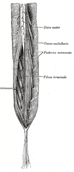
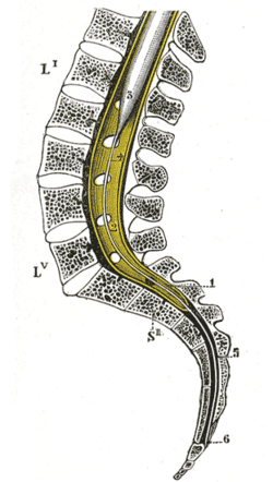
Lumbar cistern (subarachnoid space) from the conus medullaris (L1–L2) to the end of the dural sac (S2), and beyond in the sacral canal.
- Root values
Dorsal and ventral roots of L2–Co1, descending to their exit foramina.
- Origin
Lumbar, sacral and coccygeal segments of the conus medullaris.
- Course
The roots descend steeply within the dural sac, each leaving through the intervertebral foramen below its numbered vertebra (the sacral roots through the sacral foramina).
- Motor
Lower limb muscles (via the lumbar and sacral plexuses), external anal and urethral sphincters (S2–S4), pelvic floor.
- Sensory
Lower limbs, perineum and saddle area (S2–S5), bladder and rectum.
- Relations
Floats in CSF, which lets it move aside from an LP needle. Surrounded by the spinal meninges and spaces; the filum terminale runs among the roots.
Mentioned in
- Intervertebral disc
- Lumbar vertebrae
- Perineum
- Spinal cord
- Spinal meninges and spaces
- Spinal nerves and roots
Generated automatically from links in other entries.
Swipe sideways to see the whole diagram →
Swipe sideways to see the whole diagram →


Clinical correlations
Cauda equina syndrome PANCE
- Mechanism / lesion
Compression of multiple roots, most often by a large central lumbar disc herniation (L4–L5 or L5–S1); also tumor, epidural abscess or hematoma, spinal stenosis, or trauma.
- Classic presentation
Low back pain with bilateral sciatica (sometimes unilateral), saddle anesthesia, urinary retention (with overflow incontinence), fecal incontinence, reduced anal tone, sexual dysfunction, and lower motor neuron leg weakness with reduced reflexes. Emergency MRI and surgical decompression: delay worsens bladder and neurologic outcomes.
Conus medullaris syndrome (for comparison)
- Mechanism / lesion
Lesion at the conus (around L1–L2), e.g. fracture, tumor or disc.
- Classic presentation
More sudden and symmetric, with early bladder, bowel and sexual dysfunction and perianal numbness. Leg findings can be mixed UMN/LMN (e.g. brisk knee jerk with absent ankle jerk).
High-yield PANCE pearls
- Saddle anesthesia + urinary retention + bilateral sciatica + reduced anal tone → cauda equina syndrome → emergency MRI + decompression.
- Cauda equina = lower motor neuron signs (reduced reflexes, flaccid weakness).
- Check post-void residual (retention) and perianal sensation in any back pain with red flags.
Imaging anatomy
- MRI
Emergency MRI of the lumbosacral spine is the test of choice; CT myelography if MRI is unavailable or contraindicated.
- Ultrasound
Bladder scan measures post-void residual (large residual volumes support retention).
- Key views & landmarks
- Urgent MRI lumbar spine
- Bladder scan / post-void residual
Physical exam
- Surface landmarks
The cauda equina lies deep to the L2–S2 spinous processes.
- Strength / function testing
Perianal (saddle) sensation to pinprick, anal tone and voluntary squeeze on digital rectal exam, the anal wink (S2–S4) and bulbocavernosus reflexes, lower limb strength and reflexes, and post-void residual.
Self-quiz
A 45-year-old man has severe low back pain with pain radiating down both legs, numbness around the anus and inner thighs, and he hasn't urinated for 12 hours despite the urge. Anal tone is reduced. What is the most appropriate next step?
Correct answer: B
Saddle anesthesia, urinary retention, bilateral sciatica and reduced anal tone indicate cauda equina syndrome. Obtain an emergency MRI for urgent surgical decompression.
Show answer
B
Saddle anesthesia, urinary retention, bilateral sciatica and reduced anal tone indicate cauda equina syndrome. Obtain an emergency MRI for urgent surgical decompression.
What is the most common cause of cauda equina syndrome?
Correct answer: B
A large central disc herniation at L4–L5 or L5–S1 is the most common cause.
Show answer
B
A large central disc herniation at L4–L5 or L5–S1 is the most common cause.
Cauda equina compression produces which type of motor findings in the legs?
Correct answer: B
The cauda equina consists of peripheral nerve roots, so compression produces lower motor neuron signs.
Show answer
B
The cauda equina consists of peripheral nerve roots, so compression produces lower motor neuron signs.
In a patient with back pain and suspected cauda equina syndrome, which bedside test helps detect urinary retention?
Correct answer: A
Measuring the post-void residual (by bladder scan or catheter) detects urinary retention, a key feature of cauda equina syndrome, along with perianal sensation and anal tone. Suspicion warrants emergency MRI.
Show answer
A
Measuring the post-void residual (by bladder scan or catheter) detects urinary retention, a key feature of cauda equina syndrome, along with perianal sensation and anal tone. Suspicion warrants emergency MRI.
After an L1 burst fracture, a patient has sudden symmetric bladder and bowel dysfunction, perianal numbness, a brisk knee jerk and absent ankle jerks. Where is the lesion?
Correct answer: D
A conus medullaris lesion (around L1–L2) gives early, symmetric bladder, bowel and sexual dysfunction with perianal numbness and mixed UMN/LMN leg findings. Cauda equina lesions produce purely lower motor neuron signs and are often asymmetric.
Show answer
D
A conus medullaris lesion (around L1–L2) gives early, symmetric bladder, bowel and sexual dysfunction with perianal numbness and mixed UMN/LMN leg findings. Cauda equina lesions produce purely lower motor neuron signs and are often asymmetric.
Flashcards
See also
- Spinal cord
- Spinal meninges and spaces
- Intervertebral disc
- Lumbar vertebrae
- Spinal cord syndromes (comparison table)
- Back pain and foot drop (clinical case)