Triceps surae and calcaneal tendon
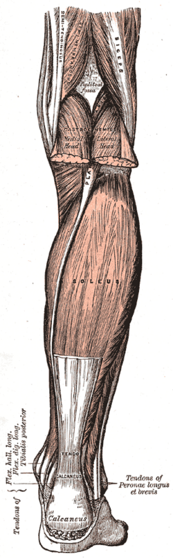
Gastrocnemius and soleus form the calf and insert via the Achilles tendon on the calcaneus to plantarflex the ankle. They carry the S1 ankle reflex; Achilles rupture and tendinopathy are common.
Core anatomy
- Location
Superficial posterior compartment of the leg (see compartments of the leg).
- Group
Posterior compartment of the leg, superficial layer
- Origin
Gastrocnemius: medial head from above the medial femoral condyle; lateral head from the lateral femoral condyle of the femur. Soleus: soleal line and posterior upper tibia and the head and posterior upper fibula. (Plantaris: small muscle above the lateral condyle with a long thin tendon.)
- Insertion
Calcaneal (Achilles) tendon into the posterior surface of the calcaneus (bones of the foot).
- Innervation
Tibial nerve (S1–S2)
- Blood supply
Sural arteries (from the popliteal artery) and the posterior tibial artery.
- Action
- Plantarflexion of the ankle (push-off in walking, running, jumping)
- Gastrocnemius also flexes the knee (it crosses the knee); it is most effective with the knee extended
- Soleus keeps the leg from falling forward when standing, and acts as a venous pump (soleal sinuses)
- Relations
The soleal venous sinuses within soleus are a common starting point for calf DVT (see deep veins of the lower limb). The sural nerve and small saphenous vein run alongside the Achilles tendon.
Found in
Mentioned in
Generated automatically from links in other entries.

Clinical correlations
Achilles tendon rupture PANCE
- Mechanism / lesion
Sudden forceful push-off or eccentric loading, typically a middle-aged "weekend warrior"; risk with fluoroquinolones and corticosteroids.
- Classic presentation
A "pop" or sensation of being kicked in the heel, then weak push-off; a palpable gap about 2–6 cm above the insertion; positive Thompson test. Patients can often still plantarflex weakly (other flexors). Treated with functional bracing or surgical repair.
Achilles tendinopathy PANCE
- Mechanism / lesion
Overuse (running), with midportion degeneration or insertional disease; fluoroquinolone association.
- Classic presentation
Morning stiffness and pain in the tendon, a thickened tender tendon about 2–6 cm above the heel; treat with eccentric calf strengthening, avoid steroid injection into the tendon.
Medial gastrocnemius strain ("tennis leg")
- Mechanism / lesion
Sudden push-off with the knee extended.
- Classic presentation
Sharp pain in the medial calf; distinguish from Achilles rupture (Thompson negative) and DVT (ultrasound).
High-yield PANCE pearls
- Pop at the heel + positive Thompson test (no plantarflexion with calf squeeze) → Achilles rupture.
- Ankle reflex = S1 (tibial nerve).
- Fluoroquinolones → tendinopathy and rupture.
- Achilles tendinopathy → eccentric strengthening; no steroid injection into the tendon.
Imaging anatomy
- X-ray
Usually normal; excludes calcaneal avulsion fracture.
- MRI
Partial vs complete tears and chronic tendinopathy.
- Ultrasound
Confirms and measures a tendon gap; shows tendinopathy (thickening, neovascularity).
- Key views & landmarks
- Dynamic ultrasound of the Achilles tendon
Physical exam
- Surface landmarks
The Achilles tendon is palpable from the calcaneus to the calf; the two gastrocnemius heads form the bulge of the upper calf.
- Palpation
Palpate along the tendon for a gap, nodularity or tenderness.
- Strength / function testing
Single-leg heel raise; ankle reflex (S1).
Thompson (Simmonds) test
- Technique
Prone with the feet over the edge of the table (or kneeling on a chair); squeeze the calf.
- Positive test
No passive plantarflexion of the foot.
- Significance
Complete Achilles tendon rupture.
Self-quiz
A 42-year-old man playing tennis feels as though someone kicked him in the back of the ankle. He can walk with a limp. When his calf is squeezed while he lies prone, the foot does not move. What is the diagnosis?
Correct answer: B
The sensation of being kicked, a limp with weak push-off, and a positive Thompson test indicate Achilles tendon rupture. Weak active plantarflexion may persist through the long toe flexors and tibialis posterior.
Show answer
B
The sensation of being kicked, a limp with weak push-off, and a positive Thompson test indicate Achilles tendon rupture. Weak active plantarflexion may persist through the long toe flexors and tibialis posterior.
The ankle jerk reflex tests which nerve root?
Correct answer: D
The ankle (Achilles) reflex is mediated by S1 via the tibial nerve to the triceps surae. The patellar reflex is L4; there is no reliable routine reflex for L5.
Show answer
D
The ankle (Achilles) reflex is mediated by S1 via the tibial nerve to the triceps surae. The patellar reflex is L4; there is no reliable routine reflex for L5.
Where do most calf deep vein thromboses begin?
Correct answer: C
DVT usually starts in the soleal venous sinuses of the calf and propagates proximally. Contraction of soleus normally squeezes these sinuses (the calf muscle pump), which immobility abolishes.
Show answer
C
DVT usually starts in the soleal venous sinuses of the calf and propagates proximally. Contraction of soleus normally squeezes these sinuses (the calf muscle pump), which immobility abolishes.
In managing Achilles tendinopathy, which treatment should be avoided?
Correct answer: D
Achilles tendinopathy is treated with eccentric calf strengthening and load management. Steroid injection into the tendon is avoided because it increases the risk of rupture. Fluoroquinolones are associated with tendinopathy and rupture.
Show answer
D
Achilles tendinopathy is treated with eccentric calf strengthening and load management. Steroid injection into the tendon is avoided because it increases the risk of rupture. Fluoroquinolones are associated with tendinopathy and rupture.
A 45-year-old tennis player feels sudden sharp pain in the medial calf while pushing off with the knee extended. The Thompson test is normal. What is the most likely diagnosis?
Correct answer: B
Tennis leg is a strain of the medial head of gastrocnemius during push-off with the knee extended. A normal Thompson test argues against Achilles rupture; ultrasound can exclude DVT if swelling suggests it.
Show answer
B
Tennis leg is a strain of the medial head of gastrocnemius during push-off with the knee extended. A normal Thompson test argues against Achilles rupture; ultrasound can exclude DVT if swelling suggests it.