Skull
The bones of the head — the neurocranium enclosing the brain and the viscerocranium forming the face. Pterion fractures, basilar skull fractures and infant fontanelles are the clinically important features.
Core anatomy
- Location
Sits on the atlas (atlas and axis (C1 and C2)) at the atlanto-occipital joints.
- Classification
Flat and irregular bones joined mainly by sutures (fibrous joints); the mandible is the only freely movable bone (see temporomandibular joint).
- Key features & landmarks
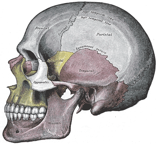
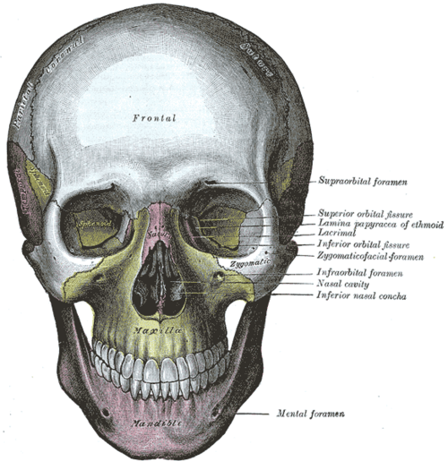
- Neurocranium (8 bones): frontal, 2 parietal, 2 temporal, occipital, sphenoid, ethmoid
- Viscerocranium (facial skeleton): 2 nasal, 2 maxillae, 2 zygomatic, 2 lacrimal, 2 palatine, 2 inferior nasal conchae, vomer and the mandible
- Sutures: coronal (frontal–parietal), sagittal (between the parietals), lambdoid (parietal–occipital), squamous (temporal–parietal)
- Pterion: H-shaped junction of the frontal, parietal, temporal and greater wing of the sphenoid, about 3–4 cm above the zygomatic arch; the thinnest part of the lateral skull, overlying the anterior branch of the middle meningeal artery
- Cranial fossae: anterior (frontal lobes; cribriform plate of the ethmoid), middle (temporal lobes, pituitary in the sella turcica; foramina rotundum, ovale, spinosum), posterior (cerebellum, brainstem; foramen magnum, internal acoustic meatus, jugular foramen, hypoglossal canal)
- Key foramina: optic canal (CN II, ophthalmic artery); superior orbital fissure (CN III, IV, V1, VI); rotundum (V2); ovale (V3); spinosum (middle meningeal artery); jugular foramen (CN IX, X, XI, internal jugular vein); hypoglossal canal (CN XII); foramen magnum (medulla, vertebral arteries)
- Relations
The dural venous sinuses groove the inner surface of the vault; the middle meningeal vessels groove the inner surface of the temporal and parietal bones.
- Ossification
The vault forms by intramembranous ossification, the base by endochondral ossification. At birth the vault bones are separated by sutures and fontanelles: the anterior fontanelle (diamond-shaped) usually closes by about 18–24 months, the posterior fontanelle (triangular) by about 2 months. verify
Muscle attachments
- Sternocleidomastoid — insertion
Joints
Generated automatically from links in other entries.
Swipe sideways to see the whole diagram →


Clinical correlations
Epidural hematoma from pterion fracture PANCE
- Mechanism / lesion
A blow to the side of the head fractures the thin pterion and tears the middle meningeal artery.
- Classic presentation
Brief loss of consciousness, a lucid interval, then rapid deterioration (headache, vomiting, ipsilateral blown pupil, contralateral hemiparesis). CT shows a biconvex (lens-shaped) hematoma that does not cross sutures. Neurosurgical emergency.
Basilar skull fracture PANCE
- Mechanism / lesion
Fracture through the skull base (often the petrous temporal bone or anterior cranial fossa) from significant head trauma.
- Classic presentation
Raccoon eyes (periorbital ecchymosis), Battle sign (mastoid ecchymosis), hemotympanum, and CSF rhinorrhea or otorrhea; possible cranial nerve palsies (VII, VIII). Avoid nasogastric tubes and nasotracheal intubation (risk of intracranial passage).
Craniosynostosis
- Mechanism / lesion
Premature fusion of one or more sutures; sagittal most common.
- Classic presentation
Abnormal head shape (e.g. long narrow scaphocephaly with sagittal synostosis); may raise intracranial pressure. Distinguish from positional plagiocephaly.
Fontanelle as a clinical sign PANCE
- Mechanism / lesion
The open anterior fontanelle reflects intracranial pressure and hydration in infants.
- Classic presentation
Bulging → raised ICP (meningitis, hydrocephalus); sunken → dehydration. Delayed closure: hypothyroidism, rickets, raised ICP; early closure: craniosynostosis.
High-yield PANCE pearls
- Lucid interval + biconvex bleed → epidural hematoma (middle meningeal artery at the pterion).
- Raccoon eyes, Battle sign, hemotympanum, CSF leak → basilar skull fracture — no NG tube.
- Bulging fontanelle = raised ICP; sunken = dehydration.
- Anterior fontanelle closes by about 18–24 months; posterior by about 2 months.
Imaging anatomy
- X-ray
Skull films are largely replaced by CT; still used in skeletal surveys for suspected child abuse.
- CT
Non-contrast head CT is the first test in head trauma (fractures with bone windows, epidural/subdural hemorrhage). CT with 3-D reconstruction for craniosynostosis and facial fractures.
- MRI
Brain parenchyma, skull base tumors and CSF leak localization (with CT cisternography).
- Ultrasound
Cranial ultrasound through the open anterior fontanelle in neonates (intraventricular hemorrhage).
- Key views & landmarks
- Non-contrast head CT (brain and bone windows)
Physical exam
- Surface landmarks
External occipital protuberance (inion), mastoid process, zygomatic arch, pterion (about two finger-breadths above the zygomatic arch and a thumb's breadth behind the frontal process of the zygomatic bone), nasion.
- Palpation
In infants, palpate the anterior fontanelle (size, tension) with the baby upright and calm; palpate sutures for ridging. In trauma, palpate for steps and bogginess.
- Strength / function testing
Inspect for raccoon eyes, Battle sign, CSF leak (clear fluid; "halo sign" on gauze) and hemotympanum on otoscopy.
Self-quiz
A 19-year-old is hit on the side of the head with a baseball. He is briefly unconscious, then talks normally for 2 hours before becoming drowsy with a dilated right pupil. CT shows a biconvex hyperdense collection. Which vessel is most likely injured?
Correct answer: B
A lucid interval with a biconvex (lentiform) extradural collection is an epidural hematoma, classically from the middle meningeal artery under the thin pterion. Subdural hematomas (crescent-shaped) come from bridging veins.
Show answer
B
A lucid interval with a biconvex (lentiform) extradural collection is an epidural hematoma, classically from the middle meningeal artery under the thin pterion. Subdural hematomas (crescent-shaped) come from bridging veins.
After a car crash, a patient has bruising around both eyes and behind the ear, and clear fluid from the nose. What should be avoided?
Correct answer: C
Raccoon eyes, Battle sign and CSF rhinorrhea indicate a basilar skull fracture. A nasogastric tube (or nasotracheal intubation) can pass through the fractured cribriform plate into the cranium; use the orogastric route.
Show answer
C
Raccoon eyes, Battle sign and CSF rhinorrhea indicate a basilar skull fracture. A nasogastric tube (or nasotracheal intubation) can pass through the fractured cribriform plate into the cranium; use the orogastric route.
Which structure passes through the foramen spinosum?
Correct answer: C
The middle meningeal artery enters the skull through the foramen spinosum. V3 passes through the foramen ovale, V2 through the foramen rotundum.
Show answer
C
The middle meningeal artery enters the skull through the foramen spinosum. V3 passes through the foramen ovale, V2 through the foramen rotundum.
After head trauma, a patient has bruising over the mastoid process. What is this sign called, and what does it suggest?
Correct answer: B
Battle sign (mastoid ecchymosis), raccoon eyes, hemotympanum and CSF rhinorrhea or otorrhea suggest a basilar skull fracture. Avoid nasogastric tubes and nasotracheal intubation.
Show answer
B
Battle sign (mastoid ecchymosis), raccoon eyes, hemotympanum and CSF rhinorrhea or otorrhea suggest a basilar skull fracture. Avoid nasogastric tubes and nasotracheal intubation.
A febrile 4-month-old has a tense, bulging anterior fontanelle while calm and upright. What does this indicate?
Correct answer: E
A bulging fontanelle indicates raised intracranial pressure (meningitis, hydrocephalus); a sunken fontanelle suggests dehydration.
Show answer
E
A bulging fontanelle indicates raised intracranial pressure (meningitis, hydrocephalus); a sunken fontanelle suggests dehydration.