Orbit
Pyramidal bony cavity holding the eyeball, extraocular muscles, nerves and vessels. Its thin floor and medial wall break in blowout fractures, and infection behind the orbital septum is an emergency.
Core anatomy
- Location
Paired cavities in the upper face, apex pointing posteromedially toward the optic canal.
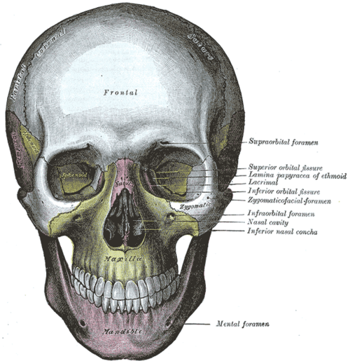
- Boundaries
- roof
- Orbital plate of the frontal bone and lesser wing of the sphenoid (anterior cranial fossa above)
- floor
- Maxilla (with zygomatic and palatine bones) — thin; the maxillary sinus lies below; infraorbital nerve in its groove
- medial wall
- Ethmoid (lamina papyracea — paper-thin), lacrimal, maxilla, sphenoid; ethmoid sinuses medially
- lateral wall
- Zygomatic bone and greater wing of the sphenoid — the thickest wall
- apex
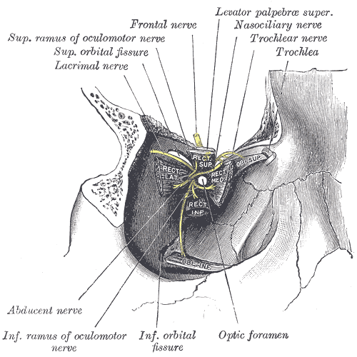
- Optic canal and superior orbital fissure
- Contents
- Eyeball and optic nerve (CN II)
- Extraocular muscles and levator palpebrae superioris
- Nerves: CN III, IV, VI, V1 branches (frontal, lacrimal, nasociliary), ciliary ganglion
- Ophthalmic artery (from the internal carotid) and superior/inferior ophthalmic veins
- Lacrimal gland (superolateral) and orbital fat
- Communications
Optic canal → middle cranial fossa (CN II, ophthalmic artery). Superior orbital fissure (CN III, IV, V1, VI, superior ophthalmic vein) → cavernous sinus (see dural venous sinuses). Inferior orbital fissure → pterygopalatine and infratemporal fossae. Ophthalmic veins are valveless and drain to the cavernous sinus — a route for spreading infection.
- Notes
The orbital septum, a fascial sheet from the orbital rim to the tarsal plates, separates the eyelids (preseptal) from the orbit proper (postseptal).
Mentioned in
- Abducens nerve (CN VI)
- Common and internal carotid arteries
- Extraocular muscles
- Eyeball
- Oculomotor nerve (CN III)
- Optic nerve and visual pathway (CN II)
- Paranasal sinuses
- Trochlear nerve (CN IV)
Generated automatically from links in other entries.
Swipe sideways to see the whole diagram →


Clinical correlations
Orbital floor ("blowout") fracture PANCE
- Mechanism / lesion
A blunt object larger than the orbit (fist, ball) raises intraorbital pressure, fracturing the thin floor (or medial wall).
- Classic presentation
Diplopia on upward gaze (entrapment of inferior rectus), enophthalmos, numbness of the cheek and upper lip (infraorbital nerve), periorbital emphysema after nose blowing. Entrapment in children can trigger the oculocardiac reflex (bradycardia, nausea) — urgent repair.
Orbital vs preseptal (periorbital) cellulitis PANCE
- Mechanism / lesion
Orbital cellulitis — infection behind the septum, usually spread from ethmoid sinusitis. Preseptal — eyelid infection in front of the septum.
- Classic presentation
Both cause eyelid redness and swelling. Red flags for orbital cellulitis: pain with eye movements, ophthalmoplegia, proptosis, decreased vision, fever. Orbital cellulitis needs CT orbits, IV antibiotics and ophthalmology; complications include abscess and cavernous sinus thrombosis.
Retrobulbar hemorrhage (orbital compartment syndrome)
- Mechanism / lesion
Bleeding behind the eye after trauma or surgery compresses the optic nerve and its blood supply.
- Classic presentation
Painful proptosis, tense orbit, reduced vision, relative afferent pupillary defect — emergency lateral canthotomy and cantholysis.
High-yield PANCE pearls
- Blowout fracture: diplopia on upgaze (inferior rectus entrapment), cheek numbness (infraorbital nerve).
- Pain with eye movement, proptosis, ophthalmoplegia → orbital cellulitis → CT + IV antibiotics.
- Orbital cellulitis usually spreads from ethmoid sinusitis.
- Ophthalmic veins → cavernous sinus (valveless) → cavernous sinus thrombosis risk.
Imaging anatomy
- X-ray
Waters view may show the "teardrop sign" of a blowout fracture, but CT has replaced it.
- CT
CT of the orbits (thin coronal slices) for fractures, muscle entrapment, orbital cellulitis and abscess.
- MRI
Optic nerve and orbital apex lesions, orbital tumors.
- Ultrasound
Ocular ultrasound for retrobulbar hematoma and globe injury (not if globe rupture is suspected with pressure).
- Key views & landmarks
- CT orbits with coronal reconstructions
Physical exam
- Surface landmarks
Orbital rim (supraorbital notch, infraorbital foramen about 1 cm below the rim), medial and lateral canthi.
- Palpation
Palpate the rim for step-offs and crepitus; test sensation of the cheek and upper lip (infraorbital nerve).
- Strength / function testing
Visual acuity, pupils (relative afferent pupillary defect), extraocular movements and diplopia, proptosis, intraocular pressure.
Self-quiz
A 22-year-old is hit in the eye by a baseball. He has double vision when looking up and numbness of the left cheek and upper lip. Which structures are most likely injured?
Correct answer: B
An orbital floor blowout fracture can trap the inferior rectus (restricting upgaze) and injure the infraorbital nerve, which runs in the floor, numbing the cheek and upper lip.
Show answer
B
An orbital floor blowout fracture can trap the inferior rectus (restricting upgaze) and injure the infraorbital nerve, which runs in the floor, numbing the cheek and upper lip.
A 7-year-old with a week of sinusitis has a red, swollen left eyelid, fever, pain when moving the eye, and limited upward gaze. What is the most appropriate next step?
Correct answer: C
Pain with eye movement and ophthalmoplegia indicate orbital (postseptal) cellulitis, usually from ethmoid sinusitis. It needs CT orbits (to exclude abscess), IV antibiotics and ophthalmology/ENT involvement.
Show answer
C
Pain with eye movement and ophthalmoplegia indicate orbital (postseptal) cellulitis, usually from ethmoid sinusitis. It needs CT orbits (to exclude abscess), IV antibiotics and ophthalmology/ENT involvement.
Which orbital wall is the thinnest?
Correct answer: C
The lamina papyracea of the ethmoid is paper-thin, which is why ethmoid sinusitis spreads to cause orbital cellulitis. The lateral wall is the thickest; the floor is also thin and fractures in blowout injuries.
Show answer
C
The lamina papyracea of the ethmoid is paper-thin, which is why ethmoid sinusitis spreads to cause orbital cellulitis. The lateral wall is the thickest; the floor is also thin and fractures in blowout injuries.
After facial trauma, a patient has painful proptosis, a tense orbit, reduced vision and a relative afferent pupillary defect. What is the most urgent intervention?
Correct answer: A
Retrobulbar hemorrhage (orbital compartment syndrome) compresses the optic nerve and its blood supply. It is an emergency treated with lateral canthotomy and cantholysis to decompress the orbit.
Show answer
A
Retrobulbar hemorrhage (orbital compartment syndrome) compresses the optic nerve and its blood supply. It is an emergency treated with lateral canthotomy and cantholysis to decompress the orbit.
Which vessel passes through the optic canal with the optic nerve?
Correct answer: B
The optic canal transmits the optic nerve (CN II) and the ophthalmic artery. The superior orbital fissure transmits CN III, IV, V1 and VI with the superior ophthalmic vein.
Show answer
B
The optic canal transmits the optic nerve (CN II) and the ophthalmic artery. The superior orbital fissure transmits CN III, IV, V1 and VI with the superior ophthalmic vein.