Inguinal triangle
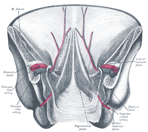
Area of the lower anterior abdominal wall, seen from inside, where direct inguinal hernias push through a weak transversalis fascia.
Core anatomy
- Location
Inferomedial part of the anterior abdominal wall, viewed from the internal (peritoneal) surface.
- Boundaries
- lateral
- Inferior epigastric vessels
- medial
- Lateral border of rectus abdominis
- inferior
- Inguinal ligament (iliopubic tract)
- floor
- Transversalis fascia (reinforced medially by the conjoint tendon)
- Contents
- No structures pass through it normally; it is an area of relative weakness in the posterior wall of the inguinal canal
- Notes
Mnemonic: "RIP" — Rectus abdominis (medial), Inferior epigastric vessels (lateral), Poupart (inguinal) ligament (inferior).
Swipe sideways to see the whole diagram →

Clinical correlations
Direct inguinal hernia PANCE
- Mechanism / lesion
Acquired weakness of the transversalis fascia (age, chronic straining, heavy lifting, chronic cough, connective tissue changes).
- Classic presentation
A medial groin bulge in an older man, usually broad-necked, easily reducible, and rarely descending into the scrotum.
High-yield PANCE pearls
- Hesselbach triangle boundaries: rectus (medial), inferior epigastrics (lateral), inguinal ligament (inferior).
- Direct hernias go through the triangle (medial to the inferior epigastrics).
Imaging anatomy
- CT
Shows a direct hernia medial to the inferior epigastric vessels.
- Ultrasound
Dynamic groin ultrasound shows a sac bulging medial to the inferior epigastric vessels on Valsalva.
Physical exam
- Surface landmarks
Lies medial to the midpoint of the inguinal ligament and just lateral to the rectus sheath, above the pubic tubercle.
- Palpation
See the groin hernia examination on inguinal canal.
Self-quiz
Which structure forms the lateral boundary of the inguinal (Hesselbach) triangle?
Correct answer: C
The inferior epigastric vessels are lateral, rectus abdominis medial, and the inguinal ligament inferior.
Show answer
C
The inferior epigastric vessels are lateral, rectus abdominis medial, and the inguinal ligament inferior.
A hernia passing through the inguinal triangle is classified as which type?
Correct answer: B
The inguinal triangle is the site of direct inguinal hernias, medial to the inferior epigastric vessels.
Show answer
B
The inguinal triangle is the site of direct inguinal hernias, medial to the inferior epigastric vessels.
Which layer is weak in a direct inguinal hernia?
Correct answer: B
The floor of the triangle is the transversalis fascia (posterior wall of the inguinal canal); its weakness allows direct herniation.
Show answer
B
The floor of the triangle is the transversalis fascia (posterior wall of the inguinal canal); its weakness allows direct herniation.
Which structure forms the medial boundary of the inguinal (Hesselbach) triangle?
Correct answer: C
"RIP": Rectus abdominis (lateral border) is medial, the Inferior epigastric vessels are lateral, and Poupart (inguinal) ligament is inferior.
Show answer
C
"RIP": Rectus abdominis (lateral border) is medial, the Inferior epigastric vessels are lateral, and Poupart (inguinal) ligament is inferior.
Compared with femoral hernias, what is characteristic of direct inguinal hernias?
Correct answer: E
Direct hernias are acquired, typically in older men, broad-necked and easily reducible, and rarely enter the scrotum, so strangulation is less likely. Femoral hernias (below the ligament, more common in women) have the highest strangulation risk; a patent processus vaginalis underlies indirect hernias.
Show answer
E
Direct hernias are acquired, typically in older men, broad-necked and easily reducible, and rarely enter the scrotum, so strangulation is less likely. Femoral hernias (below the ligament, more common in women) have the highest strangulation risk; a patent processus vaginalis underlies indirect hernias.
Flashcards
See also
- Inguinal canal
- Inferior epigastric artery
- Groin hernias (comparison table)