Latissimus dorsi
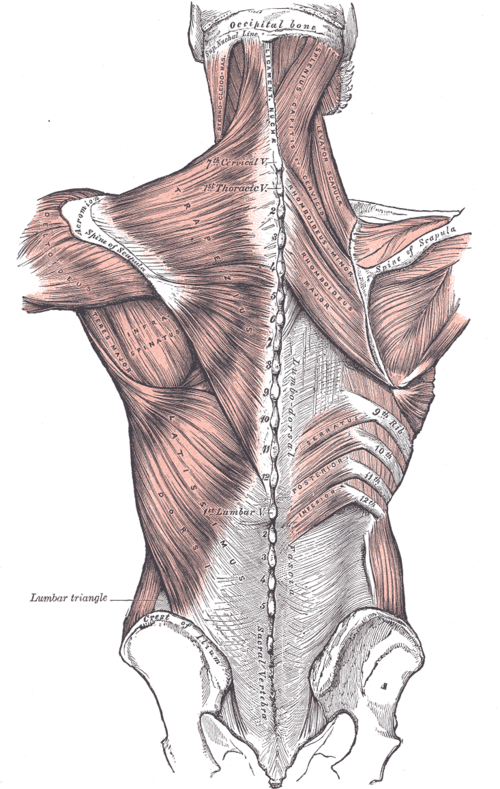
Broad flat muscle of the lower back that extends, adducts and medially rotates the arm. It is supplied by the thoracodorsal nerve and forms the posterior axillary fold.
Core anatomy
- Location
Lower and middle back, sweeping up to the axilla.
- Group
Superficial extrinsic back muscles (posterior axioappendicular).
- Origin
Spinous processes of T7–T12, the thoracolumbar fascia (and through it the lumbar and sacral spinous processes), the posterior iliac crest, the inferior 3–4 ribs, and sometimes the inferior angle of the scapula.
- Insertion
Floor of the intertubercular (bicipital) groove of the humerus.
- Innervation
Thoracodorsal nerve (C6, C7, C8), from the posterior cord of the brachial plexus.
- Blood supply
Thoracodorsal artery (from the subscapular artery) and dorsal perforators of posterior intercostal and lumbar arteries.
- Action
- Extends, adducts and medially rotates the humerus (climbing, pull-ups, swimming)
- Raises the trunk toward the arm when the arm is fixed; assists forced expiration and coughing
- Relations
With teres major it forms the posterior axillary fold. The thoracodorsal nerve and vessels run on its deep surface in the posterior wall of the axilla. Its upper border helps bound the triangle of auscultation; its lateral border helps bound the lumbar triangle.
Forms a boundary of
Generated automatically from links in other entries.

Clinical correlations
Thoracodorsal nerve injury PANCE
- Mechanism / lesion
Injury during axillary lymph node dissection (the nerve runs on the posterior axillary wall).
- Classic presentation
Weak extension, adduction and medial rotation of the arm (e.g. difficulty with climbing or pushing up from a chair). No winging.
Latissimus dorsi flap
- Mechanism / lesion
The muscle, with its thoracodorsal pedicle, is transferred for breast reconstruction after mastectomy or to cover large defects.
- Classic presentation
Usually minimal functional loss because other muscles compensate. If the thoracodorsal vessels were damaged in axillary surgery, the flap may not be possible.
High-yield PANCE pearls
- Latissimus dorsi: extension + adduction + medial rotation; thoracodorsal nerve (posterior cord).
- Axillary dissection risks: thoracodorsal (latissimus), long thoracic (serratus, winging), intercostobrachial (medial arm numbness).
- Intertubercular groove: "lady between two majors" — latissimus in the floor between pectoralis major and teres major.
Imaging anatomy
- CT
Shows muscle bulk and flap viability after reconstruction (CT angiography of the pedicle).
- MRI
Tendon avulsions (rare, in athletes such as pitchers) and muscle atrophy.
Physical exam
- Surface landmarks
Forms the posterior axillary fold with teres major.
- Palpation
Palpate the posterior axillary fold while the patient coughs or presses the arm down against resistance (contraction is felt).
- Strength / function testing
Resisted extension, adduction and medial rotation of the arm (e.g. pushing the elbow down against the examiner's hand).
Self-quiz
Latissimus dorsi inserts on which part of the humerus?
Correct answer: B
Latissimus dorsi inserts in the floor of the intertubercular groove, between pectoralis major (lateral lip) and teres major (medial lip).
Show answer
B
Latissimus dorsi inserts in the floor of the intertubercular groove, between pectoralis major (lateral lip) and teres major (medial lip).
After axillary lymph node dissection, a patient has weak extension and adduction of the arm but no scapular winging. Which nerve was injured?
Correct answer: B
The thoracodorsal nerve supplies latissimus dorsi. Long thoracic injury would cause medial winging.
Show answer
B
The thoracodorsal nerve supplies latissimus dorsi. Long thoracic injury would cause medial winging.
Which nerve roots supply latissimus dorsi through the thoracodorsal nerve?
Correct answer: C
The thoracodorsal nerve carries C6–C8 from the posterior cord.
Show answer
C
The thoracodorsal nerve carries C6–C8 from the posterior cord.
Flashcards
See also
- Axilla
- Brachial plexus
- Humerus
- Stab wound to the chest (clinical case)