Fibula
Slender lateral bone of the leg that bears little weight but forms the lateral malleolus of the ankle. The common fibular nerve wraps around its neck, where fractures and pressure injure it.
Core anatomy
- Location
Lateral bone of the leg, parallel to the tibia; not part of the knee joint but forms the lateral side of the ankle joint.
- Classification
Long bone
- Key features & landmarks
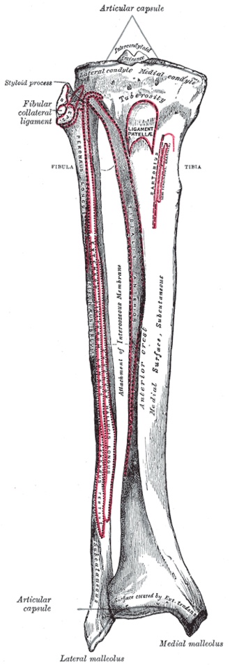
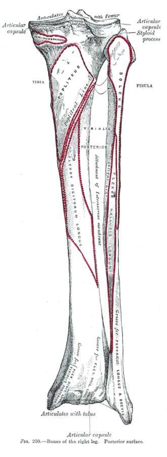
- Head: articulates with the lateral tibial condyle (proximal tibiofibular joint); attachment of biceps femoris and the fibular collateral ligament
- Neck: the common fibular nerve winds around it — palpable and vulnerable
- Shaft: slender, attachment of many leg muscles and the interosseous membrane
- Lateral malleolus: extends lower than the medial malleolus; attachment of the lateral ankle ligaments; the fibularis tendons run in a groove behind it
- Relations
The distal tibiofibular syndesmosis (anterior and posterior tibiofibular ligaments, interosseous ligament) holds the fibula to the tibia at the ankle mortise.
- Ossification
Primary center in the shaft (8th fetal week); secondary centers in the distal end (first year) and head (about 3–4 years).
Muscle attachments
- Hamstrings — insertion
- Triceps surae and calcaneal tendon — origin
Joints
Ligament attachments
Mentioned in
Generated automatically from links in other entries.


Clinical correlations
Ankle fractures (lateral malleolus) PANCE
- Mechanism / lesion
Twisting (usually supination–external rotation) injuries; Weber classification by level of the fibular fracture relative to the syndesmosis (A below, B at, C above).
- Classic presentation
Lateral ankle pain, swelling and bony tenderness at the posterior edge or tip of the lateral malleolus (Ottawa rules). Weber C and unstable (bimalleolar, widened medial clear space) fractures need fixation.
Maisonneuve fracture
- Mechanism / lesion
External rotation injury tearing the deltoid ligament or fracturing the medial malleolus, disrupting the syndesmosis, and fracturing the proximal fibula.
- Classic presentation
Medial ankle pain with proximal fibular tenderness — always palpate the whole fibula in an ankle injury.
Fibular neck fracture and common fibular nerve injury PANCE
- Mechanism / lesion
Direct blow to the lateral knee, fracture, a tight cast, prolonged leg crossing or squatting.
- Classic presentation
Foot drop (weak dorsiflexion and eversion) with numbness of the dorsum of the foot and lateral leg — see common fibular nerve.
Free fibula flap
- Mechanism / lesion
The middle fibular shaft can be harvested with the fibular artery for reconstruction (e.g. of the mandible) because the fibula is not weight-bearing.
- Classic presentation
The distal fibula is preserved to keep the ankle stable.
High-yield PANCE pearls
- Lateral malleolus tenderness → Ottawa ankle rules for X-ray.
- Medial ankle injury + proximal fibular tenderness → Maisonneuve fracture.
- Blow to the fibular neck → foot drop (common fibular nerve).
Imaging anatomy
- X-ray
Ankle AP, lateral and mortise views (Weber level, medial clear space, syndesmotic widening); full-length tibia/fibula when the proximal fibula is tender.
- CT
Complex ankle fractures and syndesmotic injury.
- MRI
Syndesmotic ligament injury (high ankle sprain) and occult fractures.
- Key views & landmarks
- Ankle mortise view (15–20° internal rotation)
Physical exam
- Surface landmarks
The fibular head is palpable on the lateral knee at about the level of the tibial tuberosity; the neck just below it; the lateral malleolus at the ankle.
- Palpation
Palpate the whole length of the fibula after an ankle injury.
Squeeze test
- Technique
Compress the tibia and fibula together at mid-calf.
- Positive test
Pain at the distal tibiofibular syndesmosis.
- Significance
Suggests a syndesmotic (high ankle) sprain.
Self-quiz
A soccer player is kicked on the lateral side of the knee. Afterward he cannot dorsiflex the right foot and has numbness over its dorsum. A fracture of which part of the fibula is most likely?
Correct answer: B
The common fibular nerve winds around the fibular neck, where a fracture or blow injures it, causing foot drop and loss of sensation on the dorsum of the foot.
Show answer
B
The common fibular nerve winds around the fibular neck, where a fracture or blow injures it, causing foot drop and loss of sensation on the dorsum of the foot.
A patient has a medial malleolar injury after an external rotation twist. Which additional exam step is essential?
Correct answer: A
A Maisonneuve fracture combines a medial ankle injury and syndesmotic disruption with a proximal fibular fracture. Palpate the whole fibula and X-ray it if tender.
Show answer
A
A Maisonneuve fracture combines a medial ankle injury and syndesmotic disruption with a proximal fibular fracture. Palpate the whole fibula and X-ray it if tender.
In the Weber classification of ankle fractures, what type is a lateral malleolus fracture above the level of the syndesmosis?
Correct answer: C
The Weber classification uses the level of the fibular fracture relative to the syndesmosis: A below, B at, C above. Weber C fractures imply syndesmotic injury and need fixation.
Show answer
C
The Weber classification uses the level of the fibular fracture relative to the syndesmosis: A below, B at, C above. Weber C fractures imply syndesmotic injury and need fixation.
Which tendons run in the groove behind the lateral malleolus?
Correct answer: E
The fibularis longus and brevis tendons pass behind the lateral malleolus (with the sural nerve and small saphenous vein). Tibialis posterior, FDL and FHL pass behind the medial malleolus.
Show answer
E
The fibularis longus and brevis tendons pass behind the lateral malleolus (with the sural nerve and small saphenous vein). Tibialis posterior, FDL and FHL pass behind the medial malleolus.
The middle of the fibular shaft can be harvested as a vascularized bone flap. Why is this possible?
Correct answer: B
The tibia carries almost all the weight through the leg, so the middle fibular shaft can be taken with the fibular artery (e.g. to reconstruct the mandible). The distal fibula is preserved because the lateral malleolus stabilizes the ankle.
Show answer
B
The tibia carries almost all the weight through the leg, so the middle fibular shaft can be taken with the fibular artery (e.g. to reconstruct the mandible). The distal fibula is preserved because the lateral malleolus stabilizes the ankle.