Eyeball
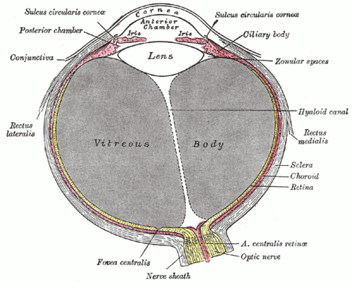
The globe of the eye — three coats (fibrous, vascular, neural) around the lens and chambers. Acute angle-closure glaucoma, retinal detachment, central retinal artery occlusion and the red eye are its emergencies.
Core anatomy
- Location
In the anterior orbit, cushioned by fat and suspended by fascia (Tenon capsule) and the suspensory ligament of Lockwood.
- Parts
- Fibrous coat: sclera (white, posterior 5/6) and cornea (transparent, avascular, anterior; most of the eye's refractive power; supplied by V1)
- Vascular coat (uvea): choroid, ciliary body (ciliary muscle for accommodation; ciliary processes secrete aqueous humor) and iris (sphincter pupillae — parasympathetic CN III; dilator pupillae — sympathetic)
- Neural coat: retina, with the macula and fovea (central vision, cones) and the optic disc (blind spot)
- Lens: suspended by zonular fibers; changes shape for near vision
- Chambers: anterior and posterior chambers (aqueous humor) and vitreous body
- Aqueous flow: ciliary body → posterior chamber → through the pupil → anterior chamber → trabecular meshwork at the iridocorneal angle → canal of Schlemm → episcleral veins
- Relations
Moved by the extraocular muscles; the optic nerve leaves posteriorly slightly medial to the posterior pole.
- Arterial supply
Ophthalmic artery (first branch of the internal carotid; see common and internal carotid arteries) → central retinal artery (inner retina — an end artery) and ciliary arteries (choroid, which supplies the outer retina).
- Venous drainage
Central retinal vein and vortex veins → ophthalmic veins → cavernous sinus.
- Lymphatic drainage
None within the globe.
- Innervation
Sensory from V1 (long and short ciliary nerves). Parasympathetic (CN III → ciliary ganglion) — pupillary constriction and accommodation. Sympathetic (superior cervical ganglion) — pupillary dilation.
Found in
Mentioned in
Generated automatically from links in other entries.

Clinical correlations
Acute angle-closure glaucoma PANCE
- Mechanism / lesion
The iris blocks the iridocorneal angle, suddenly obstructing aqueous outflow; triggered by pupil dilation (dim light, anticholinergics, mydriatics) in hyperopic older patients.
- Classic presentation
Sudden severe eye pain, headache, nausea/vomiting, blurred vision with halos; red eye, fixed mid-dilated pupil, cloudy (steamy) cornea, rock-hard eye (IOP often over 40 mm Hg). Emergency — topical timolol, pilocarpine, apraclonidine, IV/oral acetazolamide, then laser iridotomy. verify
Open-angle glaucoma PANCE
- Mechanism / lesion
Chronic optic neuropathy with increased cup-to-disc ratio, often with raised IOP; painless.
- Classic presentation
Gradual peripheral vision loss; treat with prostaglandin analog drops (latanoprost) or beta-blocker drops, laser or surgery.
Retinal detachment PANCE
- Mechanism / lesion
Separation of the neural retina from the pigment epithelium, usually from a retinal tear (myopia, trauma, older age).
- Classic presentation
Flashes (photopsia), new floaters and a "curtain" or shadow over the visual field — urgent ophthalmology.
Central retinal artery occlusion PANCE
- Mechanism / lesion
Embolus (carotid plaque, cardiac) or giant cell arteritis blocking the end artery to the inner retina.
- Classic presentation
Sudden painless monocular vision loss; cherry-red spot with a pale retina on fundoscopy. Evaluate as a stroke equivalent (carotid imaging, ESR/CRP for GCA).
The red eye PANCE
- Mechanism / lesion
Conjunctivitis (viral, bacterial, allergic), subconjunctival hemorrhage, keratitis, uveitis, acute angle closure, scleritis.
- Classic presentation
Red flags: pain, reduced vision, photophobia, abnormal pupil, corneal opacity, contact lens wear → same-day ophthalmology. Conjunctivitis has normal vision and no pain beyond grittiness.
High-yield PANCE pearls
- Red painful eye, halos, mid-dilated fixed pupil, hard eye → acute angle-closure glaucoma (emergency).
- Flashes + floaters + curtain → retinal detachment.
- Sudden painless vision loss + cherry-red spot → CRAO (stroke equivalent).
- Open-angle glaucoma: painless peripheral loss, increased cup-to-disc ratio.
- Anticholinergic or mydriatic drops can precipitate angle closure.
Imaging anatomy
- CT
Orbital CT for trauma and intraocular foreign bodies (MRI is contraindicated with metallic foreign bodies).
- MRI
Optic nerve and orbital lesions.
- Ultrasound
Ocular (B-scan) ultrasound detects retinal detachment, vitreous hemorrhage and foreign bodies when the fundus can't be seen; optic nerve sheath diameter as a marker of raised ICP.
- Key views & landmarks
- Bedside ocular ultrasound
Physical exam
- Surface landmarks
The corneoscleral junction (limbus); the optic disc lies slightly nasal to the center of the fundus.
- Palpation
Digital palpation of IOP is crude (a rock-hard eye in angle closure); tonometry measures it (normal about 10–21 mm Hg).
- Strength / function testing
Visual acuity first (Snellen), pupils (direct, consensual, swinging-flashlight for RAPD), visual fields by confrontation, extraocular movements, fundoscopy (red reflex, disc, vessels, macula), fluorescein staining for corneal abrasions.
Self-quiz
A 65-year-old woman develops severe right eye pain, blurred vision with halos around lights, and vomiting after watching a movie in a dark theater. The right eye is red with a hazy cornea and a mid-dilated pupil that does not react. What is the most likely diagnosis?
Correct answer: B
Dim light dilates the pupil, which can close a narrow iridocorneal angle. Acute angle-closure glaucoma causes a painful red eye, halos, a steamy cornea and fixed mid-dilated pupil — an emergency to lower IOP.
Show answer
B
Dim light dilates the pupil, which can close a narrow iridocorneal angle. Acute angle-closure glaucoma causes a painful red eye, halos, a steamy cornea and fixed mid-dilated pupil — an emergency to lower IOP.
A 70-year-old man with atrial fibrillation suddenly loses vision in his left eye without pain. Fundoscopy shows a pale retina with a cherry-red spot at the macula. Which vessel is occluded?
Correct answer: B
Central retinal artery occlusion makes the inner retina pale and ischemic; the thin fovea lets the underlying choroidal circulation show through as a cherry-red spot. It is a stroke equivalent.
Show answer
B
Central retinal artery occlusion makes the inner retina pale and ischemic; the thin fovea lets the underlying choroidal circulation show through as a cherry-red spot. It is a stroke equivalent.
A 58-year-old with high myopia sees sudden flashes of light, many new floaters, and a shadow spreading across part of the visual field. What is the most likely diagnosis?
Correct answer: A
Flashes (photopsia), new floaters and a curtain or shadow over the field indicate retinal detachment (risk factors: myopia, trauma, older age), which needs urgent ophthalmology.
Show answer
A
Flashes (photopsia), new floaters and a curtain or shadow over the field indicate retinal detachment (risk factors: myopia, trauma, older age), which needs urgent ophthalmology.
Through which structure does aqueous humor normally leave the anterior chamber?
Correct answer: C
Aqueous is secreted by the ciliary processes into the posterior chamber, flows through the pupil into the anterior chamber, and drains through the trabecular meshwork at the iridocorneal angle into the canal of Schlemm. Blocking the angle causes angle-closure glaucoma.
Show answer
C
Aqueous is secreted by the ciliary processes into the posterior chamber, flows through the pupil into the anterior chamber, and drains through the trabecular meshwork at the iridocorneal angle into the canal of Schlemm. Blocking the angle causes angle-closure glaucoma.
What is a common first-line medical treatment for primary open-angle glaucoma?
Correct answer: D
Open-angle glaucoma is a painless, chronic optic neuropathy with peripheral field loss. It is commonly treated with prostaglandin analog or beta-blocker drops, then laser or surgery. Mydriatics can precipitate angle closure.
Show answer
D
Open-angle glaucoma is a painless, chronic optic neuropathy with peripheral field loss. It is commonly treated with prostaglandin analog or beta-blocker drops, then laser or surgery. Mydriatics can precipitate angle closure.