Scalp
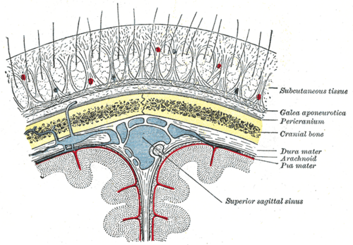
Five-layered soft tissue covering the skull vault. Its rich blood supply makes lacerations bleed heavily, and its loose areolar "danger layer" lets infection spread into the skull.
Core anatomy
- Location
Over the cranial vault from the eyebrows to the superior nuchal lines, and laterally to the zygomatic arches.
- Parts
- S — Skin: thick, hair-bearing, with many sebaceous glands
- C — Connective tissue (dense): contains the vessels and nerves; fibrous septa hold vessel walls open, so cut vessels gape and bleed profusely
- A — Aponeurosis (galea aponeurotica): joins the frontal and occipital bellies of occipitofrontalis
- L — Loose areolar tissue: the "danger layer"; allows the upper three layers to move, and blood or pus spreads freely within it (subgaleal hemorrhage)
- P — Pericranium: periosteum of the skull, attached at the sutures (limits cephalohematoma to one bone)
- Relations
The first three layers are firmly bound together and move as one ("scalp proper").
- Arterial supply
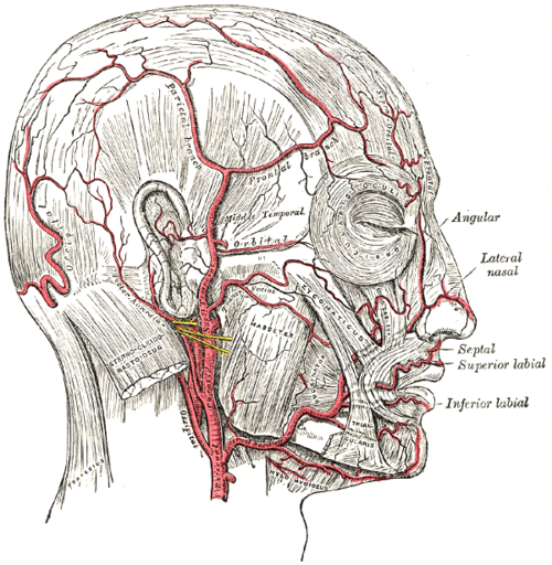
Branches of the external carotid (superficial temporal, posterior auricular, occipital) and the internal carotid via the ophthalmic artery (supratrochlear, supraorbital); extensive anastomoses.
- Venous drainage
Veins accompanying the arteries; emissary veins in the danger layer connect scalp veins with the intracranial dural venous sinuses.
- Lymphatic drainage
Occipital, posterior auricular (mastoid), preauricular (parotid) and submandibular nodes (see cervical lymph nodes).
- Innervation
Anteriorly branches of the trigeminal nerve (supratrochlear, supraorbital, zygomaticotemporal, auriculotemporal); posteriorly the greater occipital nerve (C2) (greater occipital nerve), lesser occipital and great auricular nerves (cervical plexus) and third occipital nerve.
Mentioned in
Generated automatically from links in other entries.


Clinical correlations
Scalp lacerations PANCE
- Mechanism / lesion
The dense connective tissue holds vessels open.
- Classic presentation
Heavy bleeding (significant blood loss possible in children and anticoagulated patients). Control with direct pressure and closure; repair a gaping galea to restore frontalis function and hemostasis.
Subgaleal hemorrhage and cephalohematoma PANCE
- Mechanism / lesion
Subgaleal bleeding (in the loose areolar layer), e.g. after vacuum-assisted delivery, can spread over the whole vault; cephalohematoma is subperiosteal, so it is limited by suture lines.
- Classic presentation
Subgaleal hemorrhage — a fluctuant swelling crossing sutures that can cause hypovolemic shock in newborns. Cephalohematoma — a firm swelling over one bone appearing hours after birth, risk of jaundice. Caput succedaneum — scalp edema crossing sutures, present at birth, resolves in days.
Spread of infection via emissary veins
- Mechanism / lesion
Infection in the danger layer can travel through valveless emissary veins to the dural venous sinuses.
- Classic presentation
Meningitis or venous sinus thrombosis after scalp infection (rare).
High-yield PANCE pearls
- Scalp layers: Skin, Connective tissue, Aponeurosis, Loose areolar, Pericranium.
- Cephalohematoma doesn't cross sutures; caput succedaneum and subgaleal hemorrhage do.
- Subgaleal hemorrhage in a newborn can cause shock.
Imaging anatomy
- CT
Head CT shows subgaleal hematoma and underlying fractures after trauma.
- Ultrasound
Distinguishes scalp collections in neonates.
Physical exam
- Palpation
In a newborn, a swelling limited to one bone and not crossing sutures = cephalohematoma; a boggy swelling crossing sutures = caput or subgaleal hemorrhage (monitor hemoglobin and head circumference).
- Strength / function testing
Explore lacerations for galeal tears and underlying fracture.
Self-quiz
A newborn delivered by vacuum extraction develops a boggy, fluctuant scalp swelling that crosses suture lines and increases over several hours, with tachycardia and pallor. Which layer contains the bleeding?
Correct answer: C
Subgaleal hemorrhage lies in the loose areolar tissue beneath the galea, which is not limited by sutures and can hold a large volume of blood, causing shock. A cephalohematoma is subperiosteal and limited by sutures.
Show answer
C
Subgaleal hemorrhage lies in the loose areolar tissue beneath the galea, which is not limited by sutures and can hold a large volume of blood, causing shock. A cephalohematoma is subperiosteal and limited by sutures.
Why do scalp lacerations bleed so heavily?
Correct answer: B
In the dense connective tissue layer, vessel walls are attached to fibrous septa, so cut vessels can't retract and constrict — they gape and bleed profusely.
Show answer
B
In the dense connective tissue layer, vessel walls are attached to fibrous septa, so cut vessels can't retract and constrict — they gape and bleed profusely.
A newborn has a firm scalp swelling over one parietal bone that stops sharply at the sutures. Which lesion is this?
Correct answer: C
A cephalohematoma is subperiosteal, and the pericranium is attached at the sutures, so it is limited to one bone. Caput succedaneum and subgaleal hemorrhage both cross suture lines.
Show answer
C
A cephalohematoma is subperiosteal, and the pericranium is attached at the sutures, so it is limited to one bone. Caput succedaneum and subgaleal hemorrhage both cross suture lines.