Scapula
Triangular flat bone on the posterolateral thorax that gives attachment to the rotator cuff and forms the glenoid cavity of the shoulder joint.
Core anatomy
- Location
Posterolateral thoracic wall, overlying ribs 2–7. verify
- Classification
Flat bone.
- Key features & landmarks
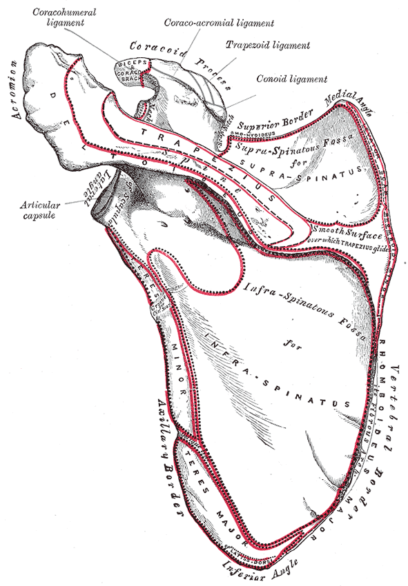
- Spine: ridge on the posterior surface, separating the supraspinous and infraspinous fossae; continues laterally as the acromion
- Acromion: articulates with the clavicle at the AC joint; forms the point of the shoulder
- Coracoid process: anterior beak-like projection (attachments: pectoralis minor, short head of biceps, coracobrachialis, coracoclavicular and coracoacromial ligaments)
- Glenoid cavity: shallow articular fossa for the humeral head
- Supraglenoid tubercle (long head of biceps brachii) and infraglenoid tubercle (long head of triceps brachii)
- Suprascapular notch: bridged by the superior transverse scapular ligament. The suprascapular nerve passes below and the artery above the ligament
- Spinoglenoid notch: between the spine and the glenoid; the suprascapular nerve passes to infraspinatus
- Fossae: supraspinous, infraspinous (posterior), subscapular (anterior/costal)
- Superior, inferior and lateral angles; medial, lateral and superior borders
- Relations
Moves on the thoracic wall at the "scapulothoracic joint" (a physiological, not anatomical, joint), held in place by serratus anterior, trapezius, the rhomboids and levator scapulae. Seventeen muscles attach to it. verify
Muscle attachments
- Biceps brachii — origin
- Deltoid — origin
- Infraspinatus — origin
- Levator scapulae — insertion
- Rhomboid major and minor — insertion
- Serratus anterior — insertion
- Subscapularis — origin
- Supraspinatus — origin
- Teres minor — origin
- Trapezius — insertion
- Triceps brachii — origin
Joints
Forms a boundary of
Mentioned in
Generated automatically from links in other entries.

Clinical correlations
Scapular fracture
- Mechanism / lesion
Uncommon. Requires high-energy trauma (motor vehicle collisions, falls from height), because the bone is mobile and protected by muscle.
- Classic presentation
Posterior shoulder pain. The main concern is associated injuries: rib fractures, pneumothorax, pulmonary contusion, and injuries to the head, spine or great vessels.
Winged scapula PANCE
- Mechanism / lesion
Medial winging: serratus anterior paralysis (long thoracic nerve). Lateral winging: trapezius paralysis (spinal accessory nerve, e.g. after posterior triangle lymph node biopsy) or rhomboid weakness (dorsal scapular nerve).
- Classic presentation
The scapular border lifts from the thoracic wall, with shoulder weakness and pain on elevating the arm.
Glenoid rim fracture (bony Bankart lesion)
- Mechanism / lesion
Anterior dislocation of the glenohumeral joint shears off the anteroinferior glenoid rim.
- Classic presentation
Recurrent instability. Glenoid bone loss influences surgical management.
High-yield PANCE pearls
- A scapular fracture implies high-energy trauma; look for rib fractures, pneumothorax and pulmonary contusion.
- Scapular Y view judges anterior vs posterior shoulder dislocation.
- Suprascapular notch: artery above, nerve below the superior transverse scapular ligament ("Army over the bridge, Navy under").
Imaging anatomy
- X-ray
The scapular Y view (true lateral scapula) shows the scapula as a "Y": the spine and acromion, the coracoid, and the body. The humeral head should be centered over the junction of the Y. It lies anterior (subcoracoid) in anterior dislocation and posterior in posterior dislocation. The AP (and Grashey, a true AP of the glenohumeral joint) and axillary views complete the shoulder series.
- CT
CT with 3D reconstruction characterizes scapular body and glenoid fractures and measures glenoid bone loss after instability.
- MRI
Soft-tissue injuries (labrum, cuff) and paralabral cysts at the spinoglenoid notch.
- Key views & landmarks
- Shoulder series: AP/Grashey, scapular Y, axillary
- Chest X-ray in trauma (ribs, pneumothorax)
Physical exam
- Surface landmarks
With the arm at the side, the medial end of the spine is at about the level of the T3 spinous process and the inferior angle at about T7. The acromion, spine and inferior angle are subcutaneous. The coracoid process can be felt deep in the deltopectoral triangle, just inferior to the junction of the middle and lateral thirds of the clavicle.
- Palpation
Palpate the acromion, spine, medial border, inferior angle and coracoid. Observe scapular position and symmetry at rest and with movement.
- Strength / function testing
Observe scapulohumeral rhythm during abduction and check for winging (wall push-up, resisted protraction).
Self-quiz
A 35-year-old unrestrained driver has a comminuted scapular body fracture on CT. Which associated injury is most important to evaluate for?
Correct answer: B
A scapular fracture implies high-energy trauma and is associated with rib fractures, pneumothorax, pulmonary contusion and other thoracic injuries.
Show answer
B
A scapular fracture implies high-energy trauma and is associated with rib fractures, pneumothorax, pulmonary contusion and other thoracic injuries.
On a scapular Y view, the humeral head lies anterior to the center of the Y, under the coracoid. What does this indicate?
Correct answer: C
On the scapular Y view the humeral head normally overlies the center of the Y. Anterior (subcoracoid) displacement indicates anterior dislocation.
Show answer
C
On the scapular Y view the humeral head normally overlies the center of the Y. Anterior (subcoracoid) displacement indicates anterior dislocation.
The suprascapular nerve passes through which structure?
Correct answer: B
The suprascapular nerve passes below the superior transverse scapular ligament through the suprascapular notch. The suprascapular artery usually passes above it.
Show answer
B
The suprascapular nerve passes below the superior transverse scapular ligament through the suprascapular notch. The suprascapular artery usually passes above it.
After a lymph node biopsy in the posterior triangle of the neck, a patient's shoulder droops and the scapula wings laterally. Which muscle is weak?
Correct answer: B
The spinal accessory nerve (CN XI) crosses the posterior triangle and is at risk during node biopsy. Trapezius paralysis causes shoulder droop and lateral winging. Medial winging points to serratus anterior (long thoracic nerve).
Show answer
B
The spinal accessory nerve (CN XI) crosses the posterior triangle and is at risk during node biopsy. Trapezius paralysis causes shoulder droop and lateral winging. Medial winging points to serratus anterior (long thoracic nerve).
The long head of triceps brachii arises from which part of the scapula?
Correct answer: A
The long head of triceps arises from the infraglenoid tubercle; the long head of biceps from the supraglenoid tubercle. The coracoid gives attachment to pectoralis minor, the short head of biceps and coracobrachialis.
Show answer
A
The long head of triceps arises from the infraglenoid tubercle; the long head of biceps from the supraglenoid tubercle. The coracoid gives attachment to pectoralis minor, the short head of biceps and coracobrachialis.