External carotid artery
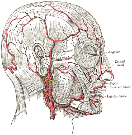
Main artery of the face, scalp and neck outside the skull. It has eight branches (SALFOPSM) and ends in the parotid gland as the superficial temporal and maxillary arteries.
Core anatomy
- Origin
Terminal branch of the common carotid artery at the upper border of the thyroid cartilage (about C3–C4).
- Course
Starts anteromedial to the internal carotid in the carotid triangle, ascends deep to the posterior belly of digastric and stylohyoid, and enters the parotid gland, where it ends behind the neck of the mandible.
- Branches
- Superior thyroid — thyroid, larynx (superior laryngeal artery), SCM (see thyroid gland)
- Ascending pharyngeal — pharynx, tonsil, meninges (see pharynx)
- Lingual — tongue, floor of mouth (see tongue)
- Facial — hooks over the mandible at the anterior border of masseter; face, tonsil, submandibular gland; ends as the angular artery
- Occipital — back of the scalp
- Posterior auricular — scalp behind the ear, auricle
- Superficial temporal (terminal) — temple and scalp (see scalp)
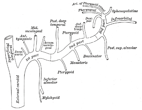
- Maxillary (terminal) — deep face: Middle meningeal artery, inferior alveolar, muscles of mastication, sphenopalatine (nasal cavity), infraorbital, palatine
- Supplies
The face, scalp, thyroid, tongue, pharynx, nasal cavity, teeth, muscles of mastication and much of the dura.
- Anastomoses
Rich anastomoses across the midline and with the internal carotid system (facial ↔ ophthalmic at the angle of the eye) — so facial and scalp wounds bleed heavily, and the ECA is a collateral path when the ICA is occluded.
- Termination
Divides into the superficial temporal and maxillary arteries within the parotid gland.
- Relations
The glossopharyngeal nerve and stylopharyngeus pass between the external and internal carotids; the hypoglossal nerve crosses superficially to both.
- Notes
Mnemonic for the branches in order — "Some Anatomists Like Freaking Out Poor Students in Medicine" (Superior thyroid, Ascending pharyngeal, Lingual, Facial, Occipital, Posterior auricular, Superficial temporal, Maxillary).
Branches
Structures supplied
- External and middle ear
- Larynx
- Muscles of facial expression
- Muscles of mastication
- Parotid gland
- Pharynx
- Sternocleidomastoid
- Submandibular gland
- Temporomandibular joint
- Thyroid gland
- Tongue
Found in
Mentioned in
Generated automatically from links in other entries.


Clinical correlations
Giant cell (temporal) arteritis PANCE
- Mechanism / lesion
Granulomatous large-vessel vasculitis affecting the superficial temporal artery and other ECA/ICA branches (ophthalmic, posterior ciliary); age over 50; linked to polymyalgia rheumatica.
- Classic presentation
New temporal headache, scalp tenderness, jaw claudication, visual loss, a tender, nodular or pulseless temporal artery, very high ESR/CRP. Start high-dose glucocorticoids immediately (before biopsy) to prevent blindness; confirm with temporal artery biopsy or ultrasound.
Epidural hematoma and posterior epistaxis PANCE
- Mechanism / lesion
Maxillary artery branches — middle meningeal artery (skull fracture at the pterion) and sphenopalatine artery (posterior nosebleeds).
- Classic presentation
See middle meningeal artery and nasal cavity. The sphenopalatine artery can be ligated or embolized for refractory posterior epistaxis.
Facial and scalp bleeding
- Mechanism / lesion
Rich blood supply and anastomoses.
- Classic presentation
Facial and scalp lacerations bleed briskly but heal well; the facial artery pulse is felt where it crosses the mandible.
High-yield PANCE pearls
- Branches = SALFOPSM; ends as superficial temporal + maxillary in the parotid.
- Age > 50, headache, jaw claudication, high ESR → giant cell arteritis → steroids first, then temporal artery biopsy.
- Maxillary artery → middle meningeal (epidural hematoma) and sphenopalatine (posterior epistaxis).
- The ECA has branches in the neck; the ICA does not.
Imaging anatomy
- CT
CTA of the head and neck.
- Ultrasound
Duplex distinguishes the ECA (branches in the neck, higher-resistance waveform) from the ICA; temporal artery ultrasound shows the "halo sign" in giant cell arteritis.
- Key views & landmarks
- Catheter angiography for embolization (epistaxis, tumors)
Physical exam
- Surface landmarks
Facial artery pulse at the lower border of the mandible at the anterior edge of masseter; superficial temporal pulse just in front of the tragus.
- Palpation
Palpate the temporal arteries for tenderness, nodularity and reduced pulse in suspected giant cell arteritis.
Self-quiz
A 72-year-old woman has a new headache over the right temple, pain in her jaw when chewing, and blurred vision. ESR is 95 mm/h. What is the most appropriate next step?
Correct answer: B
This is giant cell arteritis with visual symptoms. Start high-dose glucocorticoids immediately to prevent permanent blindness; the biopsy remains diagnostic for 1–2 weeks after starting steroids.
Show answer
B
This is giant cell arteritis with visual symptoms. Start high-dose glucocorticoids immediately to prevent permanent blindness; the biopsy remains diagnostic for 1–2 weeks after starting steroids.
Which artery is a terminal branch of the external carotid artery?
Correct answer: C
The ECA ends in the parotid gland by dividing into the maxillary and superficial temporal arteries. The ophthalmic artery is a branch of the internal carotid.
Show answer
C
The ECA ends in the parotid gland by dividing into the maxillary and superficial temporal arteries. The ophthalmic artery is a branch of the internal carotid.
An older patient on anticoagulation has heavy epistaxis flowing into the pharynx despite anterior packing. Which artery is the most likely source?
Correct answer: C
Posterior epistaxis comes from the sphenopalatine artery, a branch of the maxillary (from the external carotid). It may need posterior packing and ligation or embolization. Most anterior bleeds arise from Kiesselbach plexus.
Show answer
C
Posterior epistaxis comes from the sphenopalatine artery, a branch of the maxillary (from the external carotid). It may need posterior packing and ligation or embolization. Most anterior bleeds arise from Kiesselbach plexus.
Which cranial nerve passes between the external and internal carotid arteries?
Correct answer: A
The glossopharyngeal nerve and stylopharyngeus pass between the external and internal carotids. The hypoglossal nerve crosses superficially to both, above the bifurcation.
Show answer
A
The glossopharyngeal nerve and stylopharyngeus pass between the external and internal carotids. The hypoglossal nerve crosses superficially to both, above the bifurcation.
What is the first branch of the external carotid artery?
Correct answer: D
The branches, in order: Superior thyroid, Ascending pharyngeal, Lingual, Facial, Occipital, Posterior auricular, Superficial temporal and Maxillary. The internal carotid has no branches in the neck.
Show answer
D
The branches, in order: Superior thyroid, Ascending pharyngeal, Lingual, Facial, Occipital, Posterior auricular, Superficial temporal and Maxillary. The internal carotid has no branches in the neck.