Hepatic portal vein
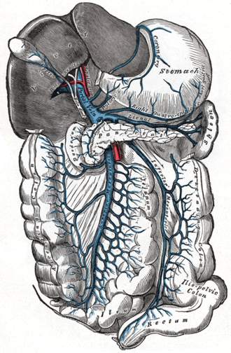
Carries nutrient-rich venous blood from the GI tract, spleen and pancreas to the liver. In portal hypertension its anastomoses with systemic veins cause varices, caput medusae and rectal varices.
Core anatomy
- Location
Formed behind the neck of the pancreas; ascends in the free edge of the hepatoduodenal ligament to the porta hepatis.
- Formation
Union of the superior mesenteric vein and the splenic vein behind the neck of the pancreas (at about L1–L2). The inferior mesenteric vein usually drains into the splenic vein (sometimes into the SMV or the confluence).
- Course
Ascends behind the 1st part of the duodenum into the hepatoduodenal ligament, where it lies posterior to the common bile duct and hepatic artery proper. At the porta hepatis it divides into right and left branches.
- Tributaries
- Superior mesenteric vein and splenic vein (formation)
- Left and right gastric veins
- Cystic veins, paraumbilical veins (via the ligamentum teres), posterior superior pancreaticoduodenal vein
- Drains into
Liver sinusoids → hepatic veins → inferior vena cava.
- Notes
Portosystemic anastomoses (where portal and systemic veins meet; dilate in portal hypertension): (1) Esophageal: left gastric (portal) ↔ esophageal veins → azygos (systemic) → esophageal varices. (2) Rectal: superior rectal (portal) ↔ middle and inferior rectal (systemic) → rectal varices (distinct from hemorrhoids). (3) Paraumbilical: paraumbilical (portal) ↔ superficial and inferior epigastric (systemic) → caput medusae. (4) Retroperitoneal: colic and other visceral veins ↔ retroperitoneal/lumbar veins. The portal system has no functional valves, so flow can reverse.
Mentioned in
Generated automatically from links in other entries.
Swipe sideways to see the whole diagram →

Clinical correlations
Portal hypertension PANCE
- Mechanism / lesion
Increased resistance to portal flow. Prehepatic: portal vein thrombosis. Intrahepatic: cirrhosis (most common), schistosomiasis. Posthepatic: Budd–Chiari, right heart failure, constrictive pericarditis.
- Classic presentation
Splenomegaly (with thrombocytopenia), ascites (SAAG ≥ 1.1 g/dL), esophageal/gastric varices (life-threatening hematemesis), caput medusae, hepatic encephalopathy.
Variceal hemorrhage PANCE
- Mechanism / lesion
Rupture of dilated esophageal (or gastric) submucosal veins at the left gastric–azygos anastomosis.
- Classic presentation
Massive hematemesis and shock in a cirrhotic patient. Resuscitate, give octreotide and prophylactic antibiotics, and perform urgent endoscopic band ligation. TIPS for refractory bleeding. Non-selective beta blockers and banding for prevention.
TIPS
- Mechanism / lesion
A transjugular intrahepatic portosystemic shunt connects a portal vein branch to a hepatic vein inside the liver, lowering portal pressure.
- Classic presentation
Used for refractory variceal bleeding or refractory ascites. Main complication is hepatic encephalopathy (portal blood bypasses the liver).
High-yield PANCE pearls
- Portal vein = SMV + splenic vein behind the neck of the pancreas.
- Anastomoses: esophageal (varices), rectal (rectal varices), paraumbilical (caput medusae), retroperitoneal.
- Variceal bleed: octreotide + antibiotics + endoscopic banding; TIPS if refractory.
- Most common cause of portal hypertension: cirrhosis.
Imaging anatomy
- CT
Contrast CT (portal venous phase) shows portal vein thrombosis, collaterals and varices, splenomegaly and ascites.
- MRI
MR venography as an alternative.
- Ultrasound
Doppler ultrasound is first-line: portal vein patency, diameter, and flow direction (normally hepatopetal, toward the liver; hepatofugal, away from the liver, in advanced portal hypertension). Also splenomegaly, ascites and recanalized paraumbilical vein.
- Key views & landmarks
- Doppler ultrasound of the portal vein
- Endoscopy (EGD) to detect and treat varices
Physical exam
- Strength / function testing
Signs of portal hypertension: caput medusae (flow away from the umbilicus), splenomegaly, ascites (shifting dullness), and a venous hum over the umbilicus (Cruveilhier–Baumgarten) in some patients. Look for stigmata of chronic liver disease.
Self-quiz
The hepatic portal vein is formed by the union of which two veins?
Correct answer: B
The superior mesenteric vein and splenic vein join behind the neck of the pancreas. The IMV usually drains into the splenic vein.
Show answer
B
The superior mesenteric vein and splenic vein join behind the neck of the pancreas. The IMV usually drains into the splenic vein.
A patient with alcohol-related cirrhosis has massive hematemesis. The bleeding varices form at an anastomosis between which veins?
Correct answer: B
Esophageal varices form where the left gastric vein (portal) meets esophageal veins that drain to the azygos (systemic). Treat with octreotide, antibiotics and endoscopic band ligation.
Show answer
B
Esophageal varices form where the left gastric vein (portal) meets esophageal veins that drain to the azygos (systemic). Treat with octreotide, antibiotics and endoscopic band ligation.
What is the most common complication after placement of a TIPS?
Correct answer: B
A TIPS diverts portal blood (with ammonia and other toxins) around the liver, so hepatic encephalopathy is the main complication.
Show answer
B
A TIPS diverts portal blood (with ammonia and other toxins) around the liver, so hepatic encephalopathy is the main complication.
Caput medusae forms at the portosystemic anastomosis between which veins?
Correct answer: B
Caput medusae is dilation of the paraumbilical veins (portal, via the ligamentum teres) anastomosing with the superficial and inferior epigastric veins (systemic). The left gastric–esophageal anastomosis forms varices, and the superior–inferior rectal anastomosis forms rectal varices.
Show answer
B
Caput medusae is dilation of the paraumbilical veins (portal, via the ligamentum teres) anastomosing with the superficial and inferior epigastric veins (systemic). The left gastric–esophageal anastomosis forms varices, and the superior–inferior rectal anastomosis forms rectal varices.
In the hepatoduodenal ligament, where does the portal vein lie relative to the common bile duct and hepatic artery?
Correct answer: E
In the free edge of the hepatoduodenal ligament, the portal vein lies posterior, with the common bile duct to the right and the hepatic artery proper to the left in front of it.
Show answer
E
In the free edge of the hepatoduodenal ligament, the portal vein lies posterior, with the common bile duct to the right and the hepatic artery proper to the left in front of it.