Tibia
The weight-bearing medial bone of the leg. Its subcutaneous shaft makes open fractures and compartment syndrome common, and the tibial tuberosity is the site of Osgood–Schlatter disease.
Core anatomy
- Location
Medial bone of the leg, from the knee joint to the ankle joint; bears almost all the body weight transmitted through the leg.
- Classification
Long bone
- Key features & landmarks
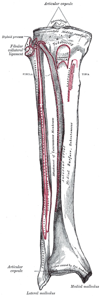
- Medial and lateral condyles (tibial plateau), separated by the intercondylar eminence (attachments of the cruciate ligaments and menisci of the knee)
- Tibial tuberosity: insertion of the patellar ligament (quadriceps)
- Gerdy tubercle on the lateral condyle: insertion of the iliotibial tract
- Shaft: triangular in cross-section; its anteromedial surface and anterior border (shin) are subcutaneous
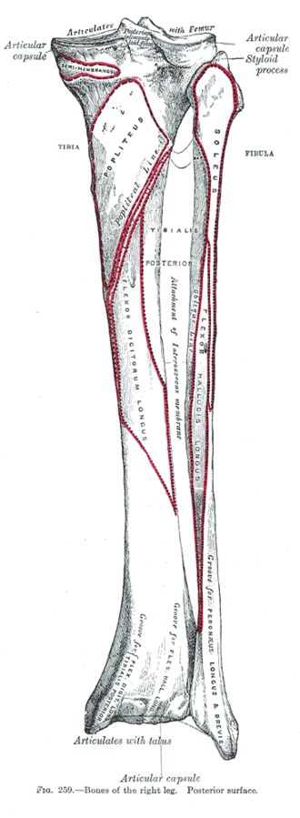
- Soleal line on the posterior surface; the interosseous border for the interosseous membrane
- Medial malleolus at the distal end; the fibular notch for the distal tibiofibular syndesmosis; the inferior articular surface (plafond) for the talus
- Relations
The anterior tibial artery and deep fibular nerve run on the interosseous membrane in the anterior compartment; the posterior tibial artery and tibial nerve lie behind it in the deep posterior compartment (see compartments of the leg). The great saphenous vein runs in front of the medial malleolus.
- Ossification
Primary center in the shaft (7th fetal week); proximal epiphysis present at or near birth; the tibial tuberosity develops as a tongue-like extension of the proximal epiphysis (the site of Osgood–Schlatter traction apophysitis).
Muscle attachments
- Quadriceps femoris — insertion
- Triceps surae and calcaneal tendon — origin
Joints
Ligament attachments
Mentioned in
Generated automatically from links in other entries.


Clinical correlations
Tibial shaft fracture and compartment syndrome PANCE
- Mechanism / lesion
Direct or twisting trauma; the most common long-bone fracture. Its subcutaneous surface makes open fractures common.
- Classic presentation
Deformity and pain; watch for acute compartment syndrome (pain out of proportion, pain on passive stretch, tense compartments) — especially the anterior compartment. Open fractures need antibiotics, tetanus prophylaxis and surgical debridement.
Osgood–Schlatter disease PANCE
- Mechanism / lesion
Traction apophysitis of the tibial tuberosity from repetitive quadriceps pull in growing adolescents (running, jumping sports).
- Classic presentation
Anterior knee pain and a tender, prominent tibial tuberosity, worse with activity and kneeling; self-limited, managed with activity modification and ice.
Tibial plateau fracture
- Mechanism / lesion
Valgus or axial load (e.g. a car bumper striking the lateral knee — "bumper fracture"), mostly the lateral plateau.
- Classic presentation
Knee effusion (lipohemarthrosis on a cross-table lateral X-ray), inability to bear weight; CT for planning; associated meniscal and ligament injuries.
Medial tibial stress syndrome and stress fracture PANCE
- Mechanism / lesion
Repetitive loading in runners and military recruits.
- Classic presentation
"Shin splints": diffuse tenderness along the posteromedial tibial border. A focal point of tenderness (especially the anterior cortex, "dreaded black line") suggests a stress fracture → MRI.
High-yield PANCE pearls
- Adolescent athlete with a tender bump at the tibial tuberosity → Osgood–Schlatter.
- Tibial fracture + pain on passive toe stretch → compartment syndrome → measure pressures/fasciotomy.
- Diffuse posteromedial shin pain = shin splints; focal tenderness = stress fracture → MRI.
- Knee X-ray fat–fluid level = lipohemarthrosis = intra-articular fracture.
Imaging anatomy
- X-ray
AP and lateral of the whole tibia including the knee and ankle. Stress fractures often don't show for 2–3 weeks.
- CT
Tibial plateau and pilon (plafond) fracture mapping.
- MRI
The most sensitive test for stress fractures and bone bruises.
- Key views & landmarks
- Full-length tibia/fibula AP and lateral
- Knee cross-table lateral for lipohemarthrosis
Physical exam
- Surface landmarks
The anterior border (shin) and anteromedial surface are subcutaneous from the tibial tuberosity to the medial malleolus; the tibial plateau margins are felt at the joint line.
- Palpation
Palpate along the shaft for focal vs diffuse tenderness; tibial tuberosity tenderness in adolescents.
- Strength / function testing
Assess the leg compartments (firmness, passive stretch pain) and distal pulses after injury.
Self-quiz
A 12-year-old boy who plays basketball has 2 months of pain below his right kneecap that is worse after games and when kneeling. There is a tender bony prominence at the tibial tuberosity. What is the most likely diagnosis?
Correct answer: B
Osgood–Schlatter disease is a traction apophysitis of the tibial tuberosity in active adolescents. It is self-limited; treatment is relative rest, ice and stretching.
Show answer
B
Osgood–Schlatter disease is a traction apophysitis of the tibial tuberosity in active adolescents. It is self-limited; treatment is relative rest, ice and stretching.
Six hours after a closed tibial shaft fracture, a patient has severe leg pain unrelieved by opioids and pain on passive plantar flexion of the toes. Pulses are palpable. What is the most likely complication?
Correct answer: B
Pain out of proportion and pain with passive stretch (here of the anterior compartment muscles) are the earliest signs of compartment syndrome. Pulses are usually preserved until late, so their presence does not exclude it.
Show answer
B
Pain out of proportion and pain with passive stretch (here of the anterior compartment muscles) are the earliest signs of compartment syndrome. Pulses are usually preserved until late, so their presence does not exclude it.
A car bumper strikes the lateral side of a pedestrian's knee. A cross-table lateral radiograph shows a fat–fluid level in the suprapatellar pouch. What injury is most likely?
Correct answer: B
A fat–fluid level (lipohemarthrosis) means marrow fat has entered the joint through an intra-articular fracture. A valgus "bumper" injury typically fractures the lateral tibial plateau; CT is used to plan treatment.
Show answer
B
A fat–fluid level (lipohemarthrosis) means marrow fat has entered the joint through an intra-articular fracture. A valgus "bumper" injury typically fractures the lateral tibial plateau; CT is used to plan treatment.
A military recruit has shin pain with running. Which finding suggests a tibial stress fracture rather than medial tibial stress syndrome ("shin splints")?
Correct answer: D
Shin splints cause diffuse tenderness along the posteromedial border. Focal point tenderness, especially over the anterior cortex, suggests a stress fracture, which may need MRI because early radiographs can be normal.
Show answer
D
Shin splints cause diffuse tenderness along the posteromedial border. Focal point tenderness, especially over the anterior cortex, suggests a stress fracture, which may need MRI because early radiographs can be normal.
Why are tibial shaft fractures so often open fractures?
Correct answer: A
The anteromedial surface and anterior border (shin) are subcutaneous, so fragments easily breach the skin. Open fractures need antibiotics, tetanus prophylaxis and surgical debridement.
Show answer
A
The anteromedial surface and anterior border (shin) are subcutaneous, so fragments easily breach the skin. Open fractures need antibiotics, tetanus prophylaxis and surgical debridement.