Spleen
Intraperitoneal lymphoid organ in the left upper quadrant under ribs 9–11. It is the organ most often injured in blunt abdominal trauma, and asplenia predisposes to infection with encapsulated bacteria.
Core anatomy
- Location
Left upper quadrant (left hypochondrium), deep to the 9th–11th ribs, with its long axis roughly along the 10th rib. Normally not palpable.
- Parts
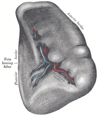
- Diaphragmatic and visceral surfaces; hilum (splenic vessels)
- Notched superior border (can help identify an enlarged spleen on exam)
- Gastrosplenic ligament (short gastric and left gastro-omental vessels) and splenorenal ligament (splenic vessels and the tail of the pancreas)
- Relations
Visceral surface contacts the stomach, left kidney, splenic (left colic) flexure and the tail of the pancreas. Separated from the ribs by the diaphragm and pleural costodiaphragmatic recess.
- Arterial supply
Splenic artery, the largest branch of the celiac trunk, running tortuously along the superior border of the pancreas.
- Venous drainage
Splenic vein, joined by the inferior mesenteric vein, unites with the superior mesenteric vein behind the neck of the pancreas to form the hepatic portal vein.
- Lymphatic drainage
Pancreaticosplenic nodes → celiac nodes.
- Notes
Functions: filters blood (removes old or abnormal RBCs and inclusions), immune response to blood-borne antigens (especially encapsulated bacteria, through IgM and opsonization), and reservoir of platelets.

Clinical correlations
Splenic injury PANCE
- Mechanism / lesion
Most commonly injured organ in blunt abdominal trauma; also lower left rib fractures. Enlarged spleens (e.g. mononucleosis) rupture more easily.
- Classic presentation
LUQ pain, Kehr sign (left shoulder pain from diaphragmatic irritation), hypotension, positive FAST. Unstable + positive FAST → laparotomy. Stable → CT grading, often non-operative management or embolization.
Splenomegaly PANCE
- Mechanism / lesion
Portal hypertension (congestion), infection (infectious mononucleosis, malaria, endocarditis), hematologic malignancy (CML, myelofibrosis, lymphoma), hemolytic anemias, infiltrative disease.
- Classic presentation
LUQ fullness, early satiety, cytopenias from hypersplenism. Mononucleosis: avoid contact sports until cleared, commonly for at least 3–4 weeks, because of rupture risk.
Asplenia and post-splenectomy infection PANCE
- Mechanism / lesion
Loss of splenic filtering and opsonization (splenectomy, sickle cell autosplenectomy).
- Classic presentation
Risk of overwhelming sepsis from encapsulated bacteria (Streptococcus pneumoniae, Haemophilus influenzae type b, Neisseria meningitidis). Howell–Jolly bodies on smear. Vaccinate against these organisms (ideally ≥ 2 weeks before elective splenectomy) and counsel about urgent care for fever.
High-yield PANCE pearls
- Blunt abdominal trauma: spleen is the most commonly injured organ; Kehr sign = left shoulder pain.
- Unstable + positive FAST → OR; stable → CT.
- Mononucleosis → splenomegaly → avoid contact sports.
- Asplenia: encapsulated organisms (pneumococcus, H. influenzae, meningococcus); vaccinate; Howell–Jolly bodies.
Imaging anatomy
- X-ray
Lower left rib fractures raise concern for splenic injury. An enlarged spleen can displace the gastric bubble and splenic flexure.
- CT
Contrast CT grades splenic injury (laceration, hematoma, active extravasation) in stable trauma patients; shows splenomegaly and infarcts.
- Ultrasound
FAST LUQ view shows free fluid around the spleen and in the splenorenal recess. Ultrasound measures splenic length; roughly more than 12–13 cm suggests splenomegaly in adults. verify
- Key views & landmarks
- FAST (LUQ view)
- CT abdomen with IV contrast (trauma)
Physical exam
- Surface landmarks
Lies under the left 9th–11th ribs posterior to the mid-axillary line. It enlarges toward the right lower quadrant (diagonally, toward the umbilicus).
- Palpation
Start palpation in the RLQ and move toward the left costal margin as the patient breathes in (so a massively enlarged spleen isn't missed). The right lateral decubitus position helps bring the spleen forward. A normal spleen is not palpable.
- Strength / function testing
Percussion for splenomegaly: Traube space (bounded by the 6th rib, the left costal margin and the anterior axillary line) is normally tympanic; dullness suggests splenomegaly. Castell sign: percuss the lowest left intercostal space in the anterior axillary line; dullness on full inspiration suggests enlargement.
Self-quiz
After a bicycle handlebar injury to the left upper abdomen, a 16-year-old is tachycardic with BP 82/50 and has left shoulder pain. FAST shows free fluid in the LUQ. What is the next step?
Correct answer: B
A hemodynamically unstable patient with a positive FAST needs laparotomy. CT is reserved for stable patients. Left shoulder pain here is Kehr sign.
Show answer
B
A hemodynamically unstable patient with a positive FAST needs laparotomy. CT is reserved for stable patients. Left shoulder pain here is Kehr sign.
A 19-year-old college athlete has fever, pharyngitis, posterior cervical lymphadenopathy and atypical lymphocytes. What advice is most important?
Correct answer: B
Infectious mononucleosis causes splenomegaly with a risk of rupture, so the patient should avoid contact sports for several weeks. Amoxicillin is avoided (it causes a rash in EBV infection).
Show answer
B
Infectious mononucleosis causes splenomegaly with a risk of rupture, so the patient should avoid contact sports for several weeks. Amoxicillin is avoided (it causes a rash in EBV infection).
Which organism is a major risk after splenectomy?
Correct answer: B
Asplenic patients are at risk from encapsulated bacteria: S. pneumoniae, H. influenzae type b and N. meningitidis. They should be vaccinated against all three.
Show answer
B
Asplenic patients are at risk from encapsulated bacteria: S. pneumoniae, H. influenzae type b and N. meningitidis. They should be vaccinated against all three.
A peripheral smear shows Howell–Jolly bodies in red cells. What do they indicate?
Correct answer: B
Howell–Jolly bodies (nuclear remnants) persist when the spleen can't filter red cells, as after splenectomy or with sickle cell autosplenectomy. These patients need vaccination against encapsulated organisms.
Show answer
B
Howell–Jolly bodies (nuclear remnants) persist when the spleen can't filter red cells, as after splenectomy or with sickle cell autosplenectomy. These patients need vaccination against encapsulated organisms.
The normal spleen lies deep to which ribs on the left?
Correct answer: D
The spleen lies deep to the 9th–11th ribs, with its long axis along the 10th rib, which is why lower left rib fractures raise concern for splenic injury. A normal spleen isn't palpable.
Show answer
D
The spleen lies deep to the 9th–11th ribs, with its long axis along the 10th rib, which is why lower left rib fractures raise concern for splenic injury. A normal spleen isn't palpable.
Flashcards
See also
- Pancreas
- Celiac trunk
- Hepatic portal vein
- Foregut, midgut and hindgut (comparison table)