Common fibular nerve
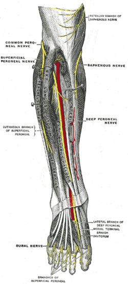
Lateral terminal branch of the sciatic nerve that winds around the fibular neck — the most commonly injured nerve of the lower limb. Injury causes foot drop and numbness of the dorsum of the foot.
Core anatomy
- Location
Lateral side of the popliteal fossa, then around the neck of the fibula into the leg.
- Root values
L4–S2.
- Origin
Sciatic nerve, at the apex of the popliteal fossa
- Course
Follows the medial border of biceps femoris to the lateral side of the popliteal fossa, passes behind the fibular head, then winds around the fibular neck (superficial, against bone) and divides deep to fibularis longus into the superficial and deep fibular nerves.
- Branches
- Superficial fibular nerve: lateral compartment (fibularis longus and brevis → eversion); skin of the lower lateral leg and most of the dorsum of the foot
- Deep fibular nerve: anterior compartment (tibialis anterior, extensor digitorum longus, extensor hallucis longus, fibularis tertius → dorsiflexion and toe extension) and extensor digitorum brevis; skin of the first web space
- Lateral sural cutaneous nerve and the sural communicating branch; articular branches to the knee
- Motor
The anterior and lateral compartments of the leg (dorsiflexion, toe extension, eversion) and short head of biceps femoris (via the sciatic division).
- Sensory
Lateral leg, dorsum of the foot, and the first web space.
- Relations
Palpable (and vulnerable) as it crosses the fibular neck just below the fibular head.
Other structures innervated
Found in
Mentioned in
Generated automatically from links in other entries.
Swipe sideways to see the whole diagram →
Swipe sideways to see the whole diagram →

Clinical correlations
Common fibular neuropathy (foot drop) PANCE
- Mechanism / lesion
Compression or trauma at the fibular neck — habitual leg crossing, prolonged squatting or kneeling, weight loss, tight casts or braces, bed rest; fibular neck fracture; knee dislocation or LCL injury.
- Classic presentation
Foot drop (weak dorsiflexion and toe extension) with weak eversion, a high-stepping (steppage) gait, and numbness of the lateral leg and dorsum of the foot. Inversion (tibial nerve) and the ankle reflex are normal — which distinguishes it from an L5 radiculopathy (inversion and hip abduction also weak).
Anterior compartment syndrome PANCE
- Mechanism / lesion
Raised pressure in the anterior compartment compresses the deep fibular nerve (see compartments of the leg).
- Classic presentation
Severe pain, pain on passive toe flexion, and early numbness of the first web space.
High-yield PANCE pearls
- Foot drop + weak eversion + dorsal foot numbness + normal inversion → common fibular nerve at the fibular neck.
- Foot drop + weak inversion and hip abduction → L5 radiculopathy.
- First web-space numbness = deep fibular nerve.
- Leg crossing, tight cast or fibular neck fracture → fibular palsy.
Imaging anatomy
- X-ray
Fibular neck fracture, knee dislocation.
- MRI
Intraneural ganglion cysts from the proximal tibiofibular joint, masses; lumbar MRI if L5 radiculopathy is suspected.
- Ultrasound
Shows compression or ganglion cysts at the fibular neck.
Physical exam
- Surface landmarks
Palpable as a cord behind the fibular head and around the fibular neck.
- Palpation
Tinel sign at the fibular neck.
- Strength / function testing
Dorsiflexion and great toe extension (deep fibular), eversion (superficial fibular), inversion (tibial — preserved), hip abduction (L5 — preserved), ankle reflex (preserved). Sensation of the first web space and dorsum of the foot. Observe for steppage gait. EMG/nerve conduction studies localize the lesion.
Self-quiz
After losing 20 kg, a woman who habitually sits with her legs crossed develops a right foot drop. Eversion is weak, inversion is normal, and the ankle reflex is intact. Sensation is reduced over the dorsum of the foot. Where is the lesion?
Correct answer: C
Weight loss (less fat padding) and leg crossing compress the common fibular nerve at the fibular neck. Weak dorsiflexion and eversion with preserved inversion (tibial nerve) and a normal ankle reflex localize the lesion there rather than to L5.
Show answer
C
Weight loss (less fat padding) and leg crossing compress the common fibular nerve at the fibular neck. Weak dorsiflexion and eversion with preserved inversion (tibial nerve) and a normal ankle reflex localize the lesion there rather than to L5.
Sensory loss limited to the first web space between the great and second toes indicates injury to which nerve?
Correct answer: B
The deep fibular nerve supplies only the skin of the first web space; the superficial fibular nerve supplies most of the dorsum of the foot.
Show answer
B
The deep fibular nerve supplies only the skin of the first web space; the superficial fibular nerve supplies most of the dorsum of the foot.
A patient has foot drop with weak ankle inversion and weak hip abduction. Where is the lesion most likely?
Correct answer: C
Foot drop with weak inversion (tibialis posterior, tibial nerve) and weak hip abduction (gluteus medius, superior gluteal nerve) points to the L5 root, which feeds several nerves. A common fibular lesion spares inversion, and a sciatic lesion in the thigh spares the hip abductors.
Show answer
C
Foot drop with weak inversion (tibialis posterior, tibial nerve) and weak hip abduction (gluteus medius, superior gluteal nerve) points to the L5 root, which feeds several nerves. A common fibular lesion spares inversion, and a sciatic lesion in the thigh spares the hip abductors.
Which gait is typical of foot drop from common fibular nerve palsy?
Correct answer: B
With weak dorsiflexion the toes drag, so the patient lifts the knee high to clear the foot: a high-stepping (steppage) gait. A Trendelenburg gait reflects hip abductor weakness.
Show answer
B
With weak dorsiflexion the toes drag, so the patient lifts the knee high to clear the foot: a high-stepping (steppage) gait. A Trendelenburg gait reflects hip abductor weakness.
A patient has weak eversion of the foot and numbness over most of the dorsum of the foot, but dorsiflexion and toe extension are normal. Which nerve is injured?
Correct answer: D
The superficial fibular nerve supplies the lateral compartment (fibularis longus and brevis → eversion) and the skin of most of the dorsum of the foot. The deep fibular nerve supplies the dorsiflexors and the first web space.
Show answer
D
The superficial fibular nerve supplies the lateral compartment (fibularis longus and brevis → eversion) and the skin of most of the dorsum of the foot. The deep fibular nerve supplies the dorsiflexors and the first web space.
Flashcards
See also
- Sciatic nerve
- Fibula
- Compartments of the leg
- Tibial nerve
- Knee ligament and meniscal injuries (comparison table)
- Lower limb nerve injuries (comparison table)
- Back pain and foot drop (clinical case)
- Severe pain under a new cast (clinical case)