Pulmonary arteries
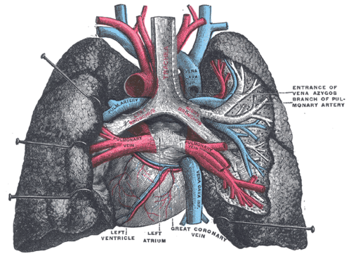
The pulmonary trunk and its right and left branches carry deoxygenated blood from the right ventricle to the lungs. They're where pulmonary emboli lodge.
Core anatomy
- Location
Middle mediastinum (pulmonary trunk within the pericardium) to the hila of the lungs.
- Origin
Right ventricle (conus arteriosus), through the pulmonary valve.
- Course
The pulmonary trunk ascends to the left of the ascending aorta and divides, at about the level of the sternal angle (T4–T5) under the aortic arch, into the right and left pulmonary arteries. The right pulmonary artery passes horizontally behind the ascending aorta and SVC to the right hilum. The left pulmonary artery is shorter, passes anterior to the descending aorta and left main bronchus, and is connected to the aortic arch by the ligamentum arteriosum.
- Branches
- Lobar and segmental arteries accompanying the bronchi into the bronchopulmonary segments
- Supplies
Alveolar capillaries for gas exchange (not the nutritive supply of the lung tissue itself, which comes from the bronchial arteries).
- Relations
At the hilum: the pulmonary artery lies anterior to the bronchus on the right and superior on the left (RALS). The ligamentum arteriosum lies between the left pulmonary artery and the arch, with the left recurrent laryngeal nerve hooking around the arch lateral to it.
- Notes
Fetal ductus arteriosus: in the fetus it shunts blood from the pulmonary trunk to the aorta, bypassing the lungs. It normally closes functionally after birth (rising oxygen, falling prostaglandins) to become the ligamentum arteriosum.
Structures supplied
Found in
Mentioned in
Generated automatically from links in other entries.

Clinical correlations
Pulmonary embolism PANCE
- Mechanism / lesion
Thromboembolus, usually from deep veins of the legs or pelvis. A large clot at the bifurcation is a saddle embolus. Risk factors (Virchow triad): stasis, hypercoagulability, endothelial injury.
- Classic presentation
Sudden dyspnea, pleuritic chest pain, tachycardia, hypoxemia, sometimes hemoptysis or syncope. Massive PE causes hypotension and RV failure. ECG most often shows sinus tachycardia; S1Q3T3 is classic but uncommon.
Patent ductus arteriosus PANCE
- Mechanism / lesion
Failure of the ductus arteriosus to close. More common in preterm infants and with congenital rubella.
- Classic presentation
Continuous "machine-like" murmur at the left infraclavicular area, with bounding pulses and wide pulse pressure. Indomethacin (or ibuprofen/acetaminophen) promotes closure; prostaglandin E1 keeps it open in ductal-dependent lesions. Large untreated PDAs can cause Eisenmenger syndrome (differential cyanosis of the lower body).
Pulmonary hypertension
- Mechanism / lesion
Elevated pulmonary arterial pressure (left heart disease, lung disease/hypoxia, chronic thromboembolism, pulmonary arterial hypertension).
- Classic presentation
Dyspnea, fatigue, a loud P2, RV heave, and signs of right heart failure. Enlarged pulmonary arteries on imaging.
High-yield PANCE pearls
- PE workup: CT pulmonary angiography; D-dimer to rule out in low/intermediate pretest probability; V/Q scan if CT contrast is contraindicated.
- PE on ECG: most common = sinus tachycardia; S1Q3T3 classic but uncommon.
- PDA: continuous machine-like murmur; indomethacin closes, PGE1 keeps open.
- Ligamentum arteriosum = remnant of the ductus arteriosus; left recurrent laryngeal nerve hooks nearby.
Imaging anatomy
- X-ray
Often normal in PE (useful to exclude other causes). Classic but uncommon signs: Hampton hump (peripheral wedge-shaped opacity from infarction) and Westermark sign (regional oligemia). Enlarged central pulmonary arteries in pulmonary hypertension.
- CT
CT pulmonary angiography (CTPA) shows filling defects in the pulmonary arteries. Also shows RV dilation (RV/LV ratio greater than 1 indicates strain) and a pulmonary trunk diameter larger than the ascending aorta (pulmonary hypertension).
- Ultrasound
Echo in massive PE: RV dilation, hypokinesis with apical sparing (McConnell sign), septal flattening (D-shaped LV), and sometimes clot in transit. Compression ultrasound of the leg veins finds the source DVT.
- Key views & landmarks
- CT pulmonary angiography
- V/Q scan (pregnancy, contrast allergy, renal failure)
Physical exam
- Surface landmarks
The pulmonary trunk lies behind the left 2nd intercostal space (pulmonic area).
- Strength / function testing
Listen for a loud P2 (pulmonary hypertension) and a continuous murmur (PDA). In suspected PE, assess vital signs, oxygen saturation, and signs of DVT.
Self-quiz
Three days after hip replacement, a 67-year-old woman has sudden dyspnea and pleuritic chest pain. HR is 118/min and SpO₂ is 89%. Renal function is normal. What is the best diagnostic test?
Correct answer: B
With a high pretest probability of PE (recent surgery, sudden hypoxic dyspnea), go straight to CTPA. A D-dimer is most useful to rule out PE when probability is low.
Show answer
B
With a high pretest probability of PE (recent surgery, sudden hypoxic dyspnea), go straight to CTPA. A D-dimer is most useful to rule out PE when probability is low.
A premature infant has a continuous machine-like murmur at the left infraclavicular area and bounding pulses. Which drug is used to close the lesion?
Correct answer: B
Patent ductus arteriosus: indomethacin (a prostaglandin synthesis inhibitor) promotes closure. Prostaglandin E1 keeps the ductus open when the circulation depends on it.
Show answer
B
Patent ductus arteriosus: indomethacin (a prostaglandin synthesis inhibitor) promotes closure. Prostaglandin E1 keeps the ductus open when the circulation depends on it.
Which structure connects the left pulmonary artery to the arch of the aorta in adults?
Correct answer: B
The ligamentum arteriosum is the fibrous remnant of the ductus arteriosus. The ligamentum venosum is the remnant of the ductus venosus; the ligamentum teres is the remnant of the umbilical vein.
Show answer
B
The ligamentum arteriosum is the fibrous remnant of the ductus arteriosus. The ligamentum venosum is the remnant of the ductus venosus; the ligamentum teres is the remnant of the umbilical vein.
The right pulmonary artery passes to the right hilum behind which structures?
Correct answer: A
The right pulmonary artery runs horizontally behind the ascending aorta and SVC. The shorter left pulmonary artery passes in front of the descending aorta and left main bronchus and is joined to the arch by the ligamentum arteriosum.
Show answer
A
The right pulmonary artery runs horizontally behind the ascending aorta and SVC. The shorter left pulmonary artery passes in front of the descending aorta and left main bronchus and is joined to the arch by the ligamentum arteriosum.
A newborn has a ductal-dependent congenital heart lesion. Which drug keeps the ductus arteriosus open until surgery?
Correct answer: E
Prostaglandin E1 keeps the ductus open in ductal-dependent lesions. Indomethacin, ibuprofen or acetaminophen (prostaglandin inhibitors) promote closure of a PDA.
Show answer
E
Prostaglandin E1 keeps the ductus open in ductal-dependent lesions. Indomethacin, ibuprofen or acetaminophen (prostaglandin inhibitors) promote closure of a PDA.