Greater omentum
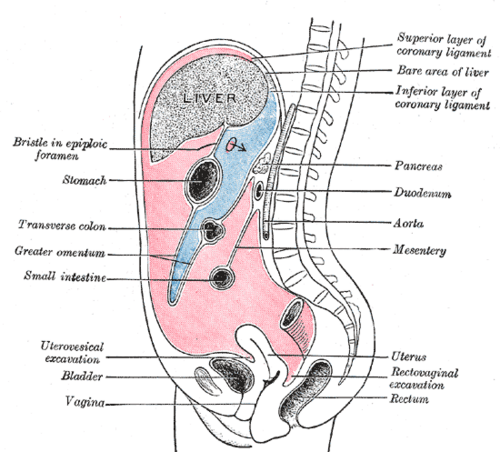
Fatty, apron-like peritoneal fold hanging from the greater curvature of the stomach. It limits the spread of infection and is a common site of peritoneal metastases.
Core anatomy
- Location
Hangs from the greater curvature of the stomach and proximal duodenum in front of the small intestine, folding back up to attach to the transverse colon.
- Parts
- Gastrocolic ligament (between the stomach and transverse colon)
- Gastrosplenic ligament (between the stomach and the spleen, containing the short gastric and left gastro-omental vessels)
- Gastrophrenic ligament
- Four layers of peritoneum (two descending, two ascending) that fuse, enclosing a potential inferior recess of the lesser sac
- Relations
Lies anterior to the small intestine and the transverse colon. The lesser omentum, by contrast, runs from the lesser curvature to the liver.
- Arterial supply
Right and left gastro-omental (gastroepiploic) arteries along the greater curvature, from the gastroduodenal and splenic arteries respectively.
- Lymphatic drainage
Contains milky spots (lymphoid aggregates) involved in peritoneal immune defense.

Clinical correlations
The "policeman of the abdomen" PANCE
- Mechanism / lesion
The mobile omentum migrates to and adheres around sites of inflammation (e.g. an inflamed appendix or perforated ulcer).
- Classic presentation
Walls off infection, forming a phlegmon or abscess rather than generalized peritonitis. Found adherent to the inflamed organ at surgery.
Omental caking (peritoneal carcinomatosis) PANCE
- Mechanism / lesion
Peritoneal spread of malignancy, classically ovarian cancer, also gastric, colorectal and pancreatic cancers.
- Classic presentation
Abdominal distension, ascites, early satiety, weight loss. Omental "cake" on imaging.
High-yield PANCE pearls
- Omental caking + ascites in a woman → consider ovarian cancer (check CA-125).
- The omentum walls off inflammation ("policeman of the abdomen").
Imaging anatomy
- CT
Normally fatty with fine vessels. Omental caking appears as soft-tissue thickening replacing the fat anterior to the bowel. Omental infarction appears as a focal fatty mass with stranding, usually in the right abdomen.
- Ultrasound
Omental caking may be seen as a hypoechoic layer beneath the abdominal wall, often with ascites.
- Key views & landmarks
- CT abdomen/pelvis with contrast
Physical exam
- Palpation
Extensive omental disease may be palpable as a firm, ill-defined upper or mid-abdominal mass with ascites.
Self-quiz
The greater omentum connects the greater curvature of the stomach to which structure?
Correct answer: B
The greater omentum hangs from the greater curvature and folds back to attach to the transverse colon (gastrocolic ligament). The lesser omentum connects the lesser curvature and duodenum to the liver.
Show answer
B
The greater omentum hangs from the greater curvature and folds back to attach to the transverse colon (gastrocolic ligament). The lesser omentum connects the lesser curvature and duodenum to the liver.
A 62-year-old woman has abdominal distension and weight loss. CT shows ascites and nodular soft tissue replacing the fat of the greater omentum. What is the most likely primary tumor?
Correct answer: B
Omental caking with ascites in a woman is classic for ovarian cancer, which spreads transcoelomically across the peritoneum. Gastric, colorectal and pancreatic cancers can also do this.
Show answer
B
Omental caking with ascites in a woman is classic for ovarian cancer, which spreads transcoelomically across the peritoneum. Gastric, colorectal and pancreatic cancers can also do this.
Which arteries run along the greater curvature of the stomach within the greater omentum?
Correct answer: B
The right and left gastro-omental (gastroepiploic) arteries run along the greater curvature. The left and right gastric arteries run along the lesser curvature.
Show answer
B
The right and left gastro-omental (gastroepiploic) arteries run along the greater curvature. The left and right gastric arteries run along the lesser curvature.