Adductor muscles of the thigh
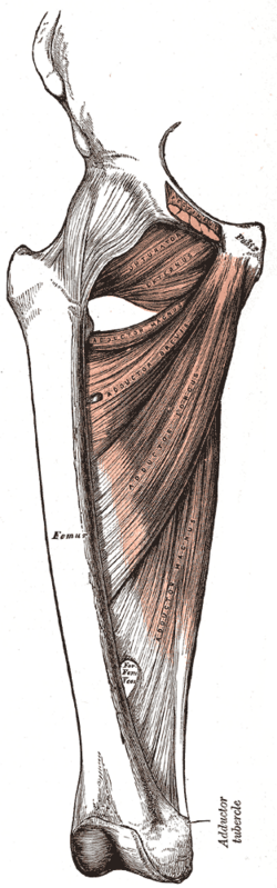
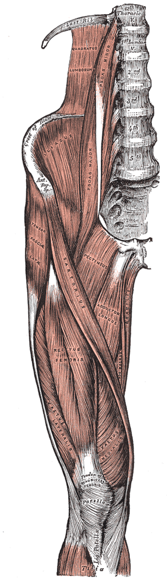
Medial compartment muscles — adductor longus, brevis and magnus, gracilis, pectineus and obturator externus — that adduct the hip. They are supplied mostly by the obturator nerve, and adductor strains are the classic "groin pull".
Core anatomy
- Location
Medial compartment of the thigh.
- Group
Medial (adductor) compartment of the thigh
- Origin
Body and inferior ramus of the pubis and the ischiopubic ramus of the hip bone; the hamstring part of adductor magnus from the ischial tuberosity.
- Insertion
The linea aspera and pectineal line of the femur; the hamstring part of adductor magnus to the adductor tubercle; gracilis to the upper medial tibia (pes anserinus).
- Innervation
Obturator nerve (L2–L4) for most; pectineus mainly by the femoral nerve; the hamstring part of adductor magnus by the tibial division of the sciatic nerve
- Blood supply
Obturator artery and branches of the deep femoral and medial circumflex femoral arteries.
- Action
- Adduction of the hip
- Assist hip flexion (longus, brevis, pectineus) or extension (hamstring part of magnus)
- Stabilize the pelvis and control lateral sway during gait
- Relations
Adductor longus forms the medial border of the femoral triangle; the adductor hiatus in adductor magnus lets the femoral vessels pass into the popliteal fossa.


Clinical correlations
Adductor (groin) strain PANCE
- Mechanism / lesion
Forceful abduction or eccentric contraction in kicking, skating and change-of-direction sports; adductor longus is most often injured.
- Classic presentation
Medial groin pain and tenderness near the pubic origin, pain on resisted adduction (squeeze test). Distinguish from hip joint pathology, osteitis pubis, inguinal hernia and athletic pubalgia.
Obturator nerve injury
- Mechanism / lesion
Pelvic surgery, pelvic fracture, childbirth, obturator hernia.
- Classic presentation
Weak adduction, a wide-based gait (circumducted), and numbness of a small area of the medial thigh.
High-yield PANCE pearls
- Groin pull in hockey/soccer players = adductor longus strain.
- Adductors = obturator nerve (except pectineus — femoral; hamstring part of magnus — tibial).
Imaging anatomy
- MRI
Adductor strains, avulsions and associated pubic bone marrow edema.
- Ultrasound
Tendon and muscle injury.
Physical exam
- Surface landmarks
The adductor longus tendon is the prominent cord in the upper medial thigh when the hip is resisted in adduction; it leads to the pubic tubercle region.
- Palpation
Tenderness at the adductor longus origin.
- Strength / function testing
Resisted adduction (squeeze test with knees flexed), passive abduction stretch.
Self-quiz
Which nerve supplies most of the adductor muscles?
Correct answer: B
The obturator nerve (L2–L4) supplies the medial compartment. Pectineus is usually supplied by the femoral nerve, and the hamstring part of adductor magnus by the tibial division of the sciatic nerve.
Show answer
B
The obturator nerve (L2–L4) supplies the medial compartment. Pectineus is usually supplied by the femoral nerve, and the hamstring part of adductor magnus by the tibial division of the sciatic nerve.
Which muscle forms the medial border of the femoral triangle?
Correct answer: B
The femoral triangle is bounded by the inguinal ligament (superior), sartorius (lateral) and the medial border of adductor longus (medial).
Show answer
B
The femoral triangle is bounded by the inguinal ligament (superior), sartorius (lateral) and the medial border of adductor longus (medial).
Through which opening do the femoral vessels pass from the thigh into the popliteal fossa?
Correct answer: D
The adductor hiatus in adductor magnus lets the femoral vessels pass into the popliteal fossa, where the femoral artery becomes the popliteal artery.
Show answer
D
The adductor hiatus in adductor magnus lets the femoral vessels pass into the popliteal fossa, where the femoral artery becomes the popliteal artery.