Facial nerve (CN VII)
Motor to the muscles of facial expression, with taste from the anterior tongue and parasympathetic supply to the lacrimal and submandibular/sublingual glands. Whether the forehead is spared separates a central (stroke) from a peripheral (Bell) palsy.
Core anatomy
- Origin
Facial motor nucleus in the caudal pons (its fibers loop around the abducens nucleus — the facial colliculus); superior salivatory nucleus (parasympathetic); nucleus of the solitary tract (taste). Emerges at the cerebellopontine angle (pontomedullary junction) as the motor root and the nervus intermedius.
- Course
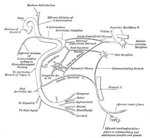
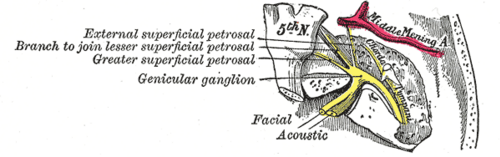
With CN VIII into the internal acoustic meatus → facial canal in the petrous temporal bone → bends at the geniculate ganglion (knee) → runs in the medial wall of the external and middle ear → descends behind it → exits the skull through the stylomastoid foramen → enters the parotid gland, where it divides into its terminal branches (it does not supply the gland).
- Branches
- Greater petrosal nerve (at the geniculate ganglion) → pterygopalatine ganglion → lacrimal gland and nasal/palatine glands
- Nerve to stapedius (damps loud sounds — lesion causes hyperacusis)
- Chorda tympani → crosses the middle ear → joins the lingual nerve (V3): taste from the anterior two-thirds of the tongue and parasympathetic to the submandibular and sublingual glands (via the submandibular ganglion)
- After the stylomastoid foramen: posterior auricular, posterior belly of digastric, stylohyoid
- Five terminal branches in the parotid: temporal, zygomatic, buccal, marginal mandibular, cervical ("To Zanzibar By Motor Car")
- Motor
Muscles of facial expression (including orbicularis oculi and oris, buccinator, platysma), stapedius, posterior belly of digastric, stylohyoid.
- Sensory
Taste from the anterior two-thirds of the tongue (chorda tympani) and a small area of skin of the external ear.
- Relations
The upper face (forehead) receives bilateral corticobulbar input, whereas the lower face receives input only from the contralateral motor cortex.
Muscles innervated
- Muscles of facial expression — Facial nerve (CN VII) — its five terminal branches leave the parotid gland: temporal, zygomatic, buccal, marginal mandibular and cervical ("To Zanzibar By Motor Car"). Also stapedius, stylohyoid and the posterior belly of digastric.
Other structures innervated
Mentioned in
Generated automatically from links in other entries.
Swipe sideways to see the whole diagram →


Clinical correlations
Bell palsy (peripheral, lower motor neuron) PANCE
- Mechanism / lesion
Idiopathic, probably viral (HSV-1) inflammation of the nerve in the facial canal. Also Lyme disease (often bilateral), Ramsay Hunt syndrome, parotid tumors, trauma, otitis media.
- Classic presentation
Acute (under 72 hours) weakness of the entire half of the face, including the forehead — cannot raise the eyebrow or close the eye (Bell phenomenon: the eye rolls up on attempted closure), drooping mouth; may have hyperacusis, loss of taste, reduced tearing, ear pain. Treat with oral corticosteroids within 72 hours (± antiviral) and eye protection (lubricants, taping at night). Most recover within weeks to months.
Central facial palsy (upper motor neuron) PANCE
- Mechanism / lesion
Stroke or tumor affecting the contralateral motor cortex or corticobulbar fibers (internal capsule).
- Classic presentation
Weakness of the contralateral lower face with forehead sparing (bilateral upper-face innervation), often with arm weakness or aphasia — a stroke until proven otherwise.
Ramsay Hunt syndrome PANCE
- Mechanism / lesion
Varicella-zoster reactivation in the geniculate ganglion.
- Classic presentation
Facial palsy with ear pain and vesicles in the external ear canal/auricle, ± hearing loss and vertigo (CN VIII). Treat with antivirals and steroids; recovery is worse than Bell palsy.
High-yield PANCE pearls
- Forehead involved → peripheral (LMN) lesion (Bell palsy); forehead spared → central (UMN) — stroke.
- Bell palsy → prednisone within 72 h ± valacyclovir + eye protection.
- Facial palsy + vesicles in the ear → Ramsay Hunt (VZV, geniculate ganglion).
- Bilateral facial palsy → Lyme disease, sarcoidosis, Guillain–Barré.
- Corneal reflex efferent = CN VII (orbicularis oculi).
- Facial palsy after parotid surgery or with a parotid mass → malignant tumor until proven otherwise.
Imaging anatomy
- CT
Temporal bone CT after trauma (fracture through the facial canal). Typical Bell palsy needs no imaging.
- MRI
MRI with contrast (internal auditory canal, temporal bone, parotid) for atypical, recurrent, slowly progressive (over 3 weeks) or non-recovering palsies — look for tumors (schwannoma, parotid malignancy).
Physical exam
- Surface landmarks
The nerve exits at the stylomastoid foramen, behind the ear lobe and anterior to the mastoid; its branches fan out from the anterior border of the parotid.
- Strength / function testing
Observe symmetry at rest (nasolabial folds, palpebral fissures). Ask the patient to raise the eyebrows (frontalis — distinguishes central from peripheral), close the eyes tightly against resistance, show the teeth, puff out the cheeks, whistle. Test taste on the anterior tongue if relevant; ask about hyperacusis and tearing. Look in the ear for vesicles.
Self-quiz
A 35-year-old woman woke with a drooping right face. She cannot wrinkle her right forehead or fully close her right eye. There are no other neurologic deficits and no ear vesicles. What is the most appropriate management?
Correct answer: B
Weakness of the whole half-face including the forehead is a peripheral (LMN) lesion — Bell palsy. Give corticosteroids within 72 hours (± antiviral) and protect the eye. Imaging is for atypical features.
Show answer
B
Weakness of the whole half-face including the forehead is a peripheral (LMN) lesion — Bell palsy. Give corticosteroids within 72 hours (± antiviral) and protect the eye. Imaging is for atypical features.
A 70-year-old man has sudden weakness of the left lower face and left arm. He can wrinkle both sides of his forehead. Where is the lesion most likely?
Correct answer: B
Forehead sparing with contralateral lower facial and arm weakness is an upper motor neuron lesion — a right hemispheric stroke (the forehead has bilateral cortical input). Activate the stroke pathway.
Show answer
B
Forehead sparing with contralateral lower facial and arm weakness is an upper motor neuron lesion — a right hemispheric stroke (the forehead has bilateral cortical input). Activate the stroke pathway.
Which nerve carries the efferent limb of the corneal reflex?
Correct answer: C
The corneal reflex has its afferent limb in V1 and its efferent limb in CN VII (orbicularis oculi closes both eyes).
Show answer
C
The corneal reflex has its afferent limb in V1 and its efferent limb in CN VII (orbicularis oculi closes both eyes).
The greater petrosal nerve, branching at the geniculate ganglion, controls which function?
Correct answer: E
The greater petrosal nerve carries parasympathetic fibers to the pterygopalatine ganglion and the lacrimal gland (and nasal and palatine glands). A lesion at or above the geniculate ganglion reduces tearing.
Show answer
E
The greater petrosal nerve carries parasympathetic fibers to the pterygopalatine ganglion and the lacrimal gland (and nasal and palatine glands). A lesion at or above the geniculate ganglion reduces tearing.
The chorda tympani carries which fibers?
Correct answer: A
The chorda tympani crosses the middle ear and joins the lingual nerve, carrying taste from the anterior two-thirds of the tongue and parasympathetic fibers to the submandibular and sublingual glands. The parotid is supplied by CN IX.
Show answer
A
The chorda tympani crosses the middle ear and joins the lingual nerve, carrying taste from the anterior two-thirds of the tongue and parasympathetic fibers to the submandibular and sublingual glands. The parotid is supplied by CN IX.