Brachial artery
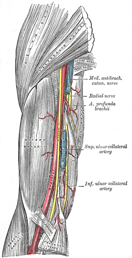
Main artery of the arm, continuing from the axillary artery. It gives the profunda brachii, then divides in the cubital fossa into the radial and ulnar arteries; it's the site for blood pressure measurement.
Core anatomy
- Location
Medial arm, then the cubital fossa.
- Origin
Continuation of the axillary artery at the inferior border of teres major.
- Course
Descends on the medial side of the arm, anterior to triceps and then brachialis, in the medial bicipital groove. The median nerve lies lateral to it proximally, crosses it (usually anteriorly) at mid-arm, and lies medial to it at the elbow. In the cubital fossa it lies medial to the biceps tendon and deep to the bicipital aponeurosis.
- Branches
- Profunda brachii (deep artery of the arm): accompanies the radial nerve in the radial groove; supplies the posterior arm and ends in collateral branches around the elbow
- Superior ulnar collateral artery: accompanies the ulnar nerve behind the medial epicondyle
- Inferior ulnar collateral artery: above the medial epicondyle
- Nutrient artery of the humerus; muscular branches
- Terminal branches: Radial artery and ulnar artery
- Supplies
Anterior arm muscles, humerus; posterior arm via the profunda brachii.
- Anastomoses
Periarticular arterial anastomosis of the elbow: the collateral arteries (from the brachial and profunda brachii) connect with the recurrent arteries (from the radial, ulnar and interosseous arteries). This network can maintain flow to the forearm if the brachial artery is slowly occluded distal to the profunda brachii.
- Termination
Divides into the radial and ulnar arteries in the cubital fossa, at about the level of the neck of the radius. A high division occurs as a variant.
- Relations
Superficial and palpable along much of its course. Accompanied by paired venae comitantes. The median cubital vein lies superficial to it, separated by the bicipital aponeurosis.
Branches
Structures supplied
Found in
Mentioned in
Generated automatically from links in other entries.
Swipe sideways to see the whole diagram →

Clinical correlations
Supracondylar fracture with arterial injury PANCE
- Mechanism / lesion
Displaced extension-type supracondylar humerus fracture in a child; the proximal fragment can tent, kink or tear the artery.
- Classic presentation
A pulseless hand that may be pink (collateral-perfused) or white (ischemic). Forearm compartment syndrome (pain out of proportion, pain with passive finger extension) can lead to Volkmann ischemic contracture if unrecognized.
Blood pressure measurement PANCE
- Mechanism / lesion
The cuff occludes the brachial artery against the humerus.
- Classic presentation
Korotkoff sounds are auscultated over the artery in the cubital fossa, medial to the biceps tendon. The cuff should fit the arm, with its bladder centered over the artery.
Proximal hemorrhage control
- Mechanism / lesion
The artery lies superficially along the medial arm and can be compressed against the humerus.
- Classic presentation
Manual compression at mid-arm, medial to the humerus, controls distal bleeding temporarily.
High-yield PANCE pearls
- Supracondylar fracture → brachial artery injury + median/AIN; watch for compartment syndrome and Volkmann contracture.
- BP measurement: auscultate the brachial artery medial to the biceps tendon in the cubital fossa.
- Profunda brachii travels with the radial nerve in the radial groove.
- Cubital fossa contents, lateral to medial: biceps tendon, brachial artery, median nerve.
Imaging anatomy
- X-ray
Not visible. Assess fractures (supracondylar) that threaten it.
- CT
CT angiography in trauma (e.g. a pulseless supracondylar fracture that stays pulseless after reduction) or suspected occlusion.
- Ultrasound
Duplex ultrasound shows flow, stenosis and pseudoaneurysm (e.g. after catheterization or arterial puncture). The artery is a landmark for ultrasound-guided access to the brachial veins.
- Key views & landmarks
- Duplex ultrasound of the brachial artery
- CT angiography of the upper extremity
Physical exam
- Surface landmarks
Pulse palpable in the medial bicipital groove of the arm, and in the cubital fossa medial to the biceps tendon (feel the tendon while the patient flexes against resistance, then move medially).
- Palpation
Palpate with the elbow slightly flexed and relaxed.
- Strength / function testing
In elbow trauma, assess the radial pulse, capillary refill, color, temperature, and pain on passive finger extension (compartment syndrome).
Self-quiz
A 5-year-old boy has a displaced supracondylar humerus fracture. Six hours after closed reduction he has severe forearm pain that worsens when his fingers are passively extended. What complication is developing?
Correct answer: B
Compartment syndrome from brachial artery injury, ischemia and swelling presents with pain out of proportion and pain on passive stretch. It needs urgent fasciotomy; untreated, it leads to Volkmann ischemic contracture.
Show answer
B
Compartment syndrome from brachial artery injury, ischemia and swelling presents with pain out of proportion and pain on passive stretch. It needs urgent fasciotomy; untreated, it leads to Volkmann ischemic contracture.
Where is the stethoscope placed to auscultate Korotkoff sounds during blood pressure measurement?
Correct answer: B
The brachial artery lies medial to the biceps tendon in the cubital fossa, with the median nerve medial to the artery.
Show answer
B
The brachial artery lies medial to the biceps tendon in the cubital fossa, with the median nerve medial to the artery.
Which branch of the brachial artery accompanies the radial nerve in the radial groove?
Correct answer: C
The profunda brachii (deep artery of the arm) runs with the radial nerve. The superior ulnar collateral artery accompanies the ulnar nerve.
Show answer
C
The profunda brachii (deep artery of the arm) runs with the radial nerve. The superior ulnar collateral artery accompanies the ulnar nerve.
At about what level does the brachial artery normally divide into the radial and ulnar arteries?
Correct answer: D
The brachial artery ends in the cubital fossa at about the level of the neck of the radius by dividing into the radial and ulnar arteries. It begins at the inferior border of teres major. A high division in the arm is a recognized variant.
Show answer
D
The brachial artery ends in the cubital fossa at about the level of the neck of the radius by dividing into the radial and ulnar arteries. It begins at the inferior border of teres major. A high division in the arm is a recognized variant.
A 6-year-old with a displaced supracondylar humerus fracture has no palpable radial pulse, but the hand is warm and pink with brisk capillary refill. What best explains the perfusion?
Correct answer: B
A pink, pulseless hand is perfused by collaterals: the collateral branches of the brachial and profunda brachii arteries connect with the recurrent branches of the radial, ulnar and interosseous arteries. It still needs urgent reduction and close monitoring; a white, pulseless hand indicates ischemia.
Show answer
B
A pink, pulseless hand is perfused by collaterals: the collateral branches of the brachial and profunda brachii arteries connect with the recurrent branches of the radial, ulnar and interosseous arteries. It still needs urgent reduction and close monitoring; a white, pulseless hand indicates ischemia.