Vertebral artery
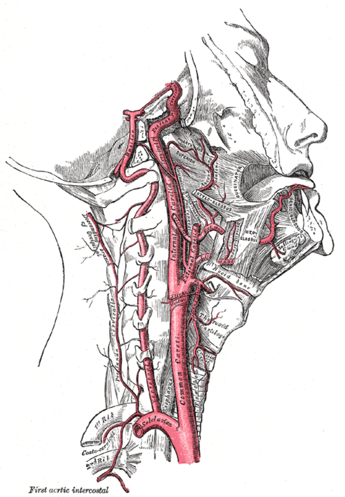
First branch of the subclavian artery, ascending through the cervical transverse foramina to join its partner and form the basilar artery. Dissection and subclavian steal are the key clinical problems.
Core anatomy
- Location
Deep neck, within the transverse foramina of the cervical vertebrae, then the suboccipital region and posterior cranial fossa.
- Origin
First part of the subclavian artery (its first branch).
- Course
V1 (pre-foraminal): ascends to enter the transverse foramen of C6 (usually). V2 (foraminal): rises through the transverse foramina of C6–C1. V3 (atlantic): curves around the lateral mass and along the posterior arch of the atlas in the suboccipital triangle. V4 (intracranial): pierces the dura, passes through the foramen magnum, and joins the other vertebral artery at the pontomedullary junction to form the basilar artery.
- Branches
- Spinal (segmental) branches in the neck
- Anterior spinal artery (the two vertebral contributions unite) and posterior spinal arteries
- Posterior inferior cerebellar artery (PICA), supplying the lateral medulla and inferior cerebellum
- Meningeal branches
- Supplies
Cervical spinal cord and vertebrae, and (via the basilar artery) the brainstem, cerebellum, occipital lobes and thalami — the posterior circulation.
- Anastomoses
Through the basilar artery and posterior communicating arteries, the circle of Willis connects with the carotid system.
- Relations
Surrounded by a sympathetic plexus in the transverse foramina. In the suboccipital triangle it lies on the posterior arch of C1.
Branches
Structures supplied
Found in
Mentioned in
Generated automatically from links in other entries.

Clinical correlations
Vertebral artery dissection PANCE
- Mechanism / lesion
An intimal tear after neck trauma, sudden neck movements or cervical manipulation, or spontaneously (connective tissue disorders).
- Classic presentation
Neck pain and occipital headache followed by posterior circulation ischemia: vertigo, ataxia, diplopia, dysarthria, dysphagia, and lateral medullary (Wallenberg) syndrome (ipsilateral facial and contralateral body pain/temperature loss, ipsilateral Horner syndrome, hoarseness). A common cause of stroke in younger adults.
Subclavian steal syndrome PANCE
- Mechanism / lesion
Stenosis of the proximal subclavian artery (before the vertebral origin) makes blood flow backward down the ipsilateral vertebral artery to supply the arm.
- Classic presentation
Arm claudication and vertebrobasilar symptoms (dizziness, syncope) with arm exercise. A systolic BP difference between the arms (commonly more than 15–20 mmHg) and a weaker pulse on the affected side. verify
Vertebral artery injury with cervical fractures
- Mechanism / lesion
Fractures involving the transverse foramina, facet dislocations, or upper cervical injuries.
- Classic presentation
Often asymptomatic initially; screening CT angiography in high-risk injury patterns.
High-yield PANCE pearls
- Vertebral artery: 1st branch of subclavian, enters C6 transverse foramen, joins at pontomedullary junction → basilar.
- Neck pain + posterior circulation stroke after neck manipulation → vertebral artery dissection.
- Arm BP difference + dizziness with arm exercise → subclavian steal (reversed vertebral flow).
- PICA occlusion → Wallenberg (lateral medullary) syndrome.
Imaging anatomy
- CT
CT angiography of the head and neck: dissection (tapered narrowing, intimal flap, pseudoaneurysm), stenosis, and injury after trauma.
- MRI
MRI/MRA with fat-suppressed sequences shows intramural hematoma (crescent sign) in dissection and posterior circulation infarcts on diffusion imaging.
- Ultrasound
Duplex ultrasound shows reversed flow in the vertebral artery in subclavian steal.
- Key views & landmarks
- CTA or MRA of the neck and head
- Duplex of the vertebral and subclavian arteries
Physical exam
- Surface landmarks
The V2 segment lies deep in the transverse foramina and can't be palpated.
- Strength / function testing
Bilateral arm blood pressures and radial pulses (subclavian steal). Cranial nerve and cerebellar exam (nystagmus, dysmetria, gait) for posterior circulation ischemia. Horner syndrome and crossed sensory loss suggest a lateral medullary infarct.
Self-quiz
A 38-year-old woman develops occipital headache and neck pain 2 days after chiropractic neck manipulation, followed by vertigo, dysarthria and a right Horner syndrome. What is the most likely diagnosis?
Correct answer: B
Neck pain after manipulation followed by posterior circulation signs and Horner syndrome (lateral medulla) suggests vertebral artery dissection. Carotid dissection causes anterior circulation (hemispheric) symptoms.
Show answer
B
Neck pain after manipulation followed by posterior circulation signs and Horner syndrome (lateral medulla) suggests vertebral artery dissection. Carotid dissection causes anterior circulation (hemispheric) symptoms.
A 64-year-old smoker becomes dizzy while painting a ceiling with his left arm. BP is 150/80 on the right and 118/70 on the left. What is the mechanism of his symptoms?
Correct answer: B
Subclavian steal: proximal subclavian stenosis causes reversed flow down the ipsilateral vertebral artery when the arm works, "stealing" from the posterior circulation.
Show answer
B
Subclavian steal: proximal subclavian stenosis causes reversed flow down the ipsilateral vertebral artery when the arm works, "stealing" from the posterior circulation.
Through which transverse foramen does the vertebral artery usually first enter the cervical spine?
Correct answer: B
It usually enters at C6 (C7's transverse foramen normally transmits only veins) and ascends through C6–C1.
Show answer
B
It usually enters at C6 (C7's transverse foramen normally transmits only veins) and ascends through C6–C1.
Which branch of the vertebral artery supplies the lateral medulla and inferior cerebellum?
Correct answer: C
PICA is a branch of the vertebral artery; its occlusion (or vertebral dissection) causes lateral medullary (Wallenberg) syndrome. AICA, SCA and PCA arise from the basilar artery.
Show answer
C
PICA is a branch of the vertebral artery; its occlusion (or vertebral dissection) causes lateral medullary (Wallenberg) syndrome. AICA, SCA and PCA arise from the basilar artery.
Where do the two vertebral arteries join to form the basilar artery?
Correct answer: B
The vertebral arteries pass through the foramen magnum and unite at the pontomedullary junction to form the basilar artery, which ends by dividing into the posterior cerebral arteries.
Show answer
B
The vertebral arteries pass through the foramen magnum and unite at the pontomedullary junction to form the basilar artery, which ends by dividing into the posterior cerebral arteries.