Ribs
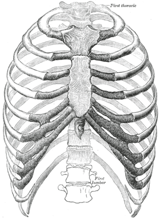
Twelve pairs of curved bones forming most of the thoracic cage. They are grouped as true, false and floating ribs, and rib fractures are the most common thoracic injury.
Core anatomy
- Location
Thoracic cage, articulating posteriorly with the thoracic vertebrae and anteriorly (via costal cartilages) with the sternum or each other.
- Classification
Flat bones.
- Key features & landmarks
- True ribs (1–7): attach directly to the sternum via their own costal cartilages (vertebrosternal)
- False ribs (8–10): cartilages join the cartilage of the rib above (vertebrochondral), forming the costal margin
- Floating ribs (11–12): no anterior attachment
- Typical rib (3–9): head with two facets (articulates with its own vertebra and the one above), neck, tubercle (articulates with the transverse process), angle, shaft, and costal groove on the inferior internal surface for the intercostal vessels and nerve
- 1st rib: broadest, shortest and most curved; scalene tubercle (anterior scalene) separates the groove for the subclavian vein (anterior) from the groove for the subclavian artery (posterior)
- Ribs 10–12: single facet on the head; 11 and 12 have no neck or tubercle
- Relations
The intercostal neurovascular bundle runs in the costal groove at the inferior border of each rib (see intercostal space).

Clinical correlations
Rib fractures PANCE
- Mechanism / lesion
Blunt chest trauma. The middle ribs are most commonly fractured, usually just anterior to the angle (the weakest point).
- Classic presentation
Point tenderness, pain with breathing and coughing, and splinting. Complications include pneumothorax, hemothorax and pulmonary contusion. In older adults, pain-limited breathing leads to atelectasis and pneumonia. Lower rib (9–12) fractures raise concern for splenic, hepatic or renal injury. 1st and 2nd rib fractures imply high-energy trauma, with possible injury to the subclavian vessels or brachial plexus.
Flail chest PANCE
- Mechanism / lesion
Fracture of three or more adjacent ribs in two or more places, creating a free-floating segment. verify
- Classic presentation
Paradoxical movement: the segment moves inward on inspiration and outward on expiration. Often accompanied by underlying pulmonary contusion, which drives the respiratory failure.
Cervical rib
- Mechanism / lesion
Supernumerary rib articulating with C7, present in a small percentage of people.
- Classic presentation
Often asymptomatic. Can compress the lower trunk of the brachial plexus or the subclavian artery (thoracic outlet syndrome).
Costochondritis PANCE
- Mechanism / lesion
Inflammation of the costochondral or costosternal junctions (Tietze syndrome if there is visible swelling).
- Classic presentation
Anterior chest wall pain reproduced by palpation of the costal cartilages. A diagnosis of exclusion after cardiac and other causes of chest pain are considered.
High-yield PANCE pearls
- Flail chest = paradoxical motion of a chest-wall segment; the real danger is the underlying pulmonary contusion.
- Lower rib fractures → look for spleen/liver/kidney injury.
- 1st rib fracture → high-energy mechanism; consider great vessel and brachial plexus injury.
- Rib notching (inferior margins, ribs ~3–8) on CXR = coarctation of the aorta (enlarged intercostal collaterals).
- Costochondritis: chest wall pain reproducible on palpation; rule out cardiac causes first.
Imaging anatomy
- X-ray
The chest X-ray is used mainly to detect complications (pneumothorax, hemothorax, contusion). It misses many rib fractures, especially anterior and costochondral ones. Dedicated rib series add sensitivity. Rib notching of the inferior rib margins suggests coarctation.
- CT
CT chest is the most sensitive test for rib fractures and for associated injuries (contusion, hemothorax, solid-organ injury).
- Ultrasound
Can show cortical disruption at a point of tenderness and detect pneumothorax and effusion.
- Key views & landmarks
- PA/AP chest radiograph
- CT chest (trauma protocol)
Physical exam
- Surface landmarks
Count ribs anteriorly from the 2nd costal cartilage at the sternal angle. The costal margin is formed by cartilages 7–10. Rib 12 is felt posteriorly; the inferior angle of the scapula (arm at the side) lies at about rib 7 or the 7th intercostal space.
- Palpation
Palpate along each rib for point tenderness, crepitus and step-off. Compress the chest anteroposteriorly and laterally (pain at a distant site suggests fracture).
- Strength / function testing
Observe chest expansion for symmetry and paradoxical movement (flail segment).
Self-quiz
After a motorcycle crash, a patient has a segment of the right chest wall that moves inward during inspiration. Imaging shows fractures of ribs 4–7, each broken in two places. Which associated condition most often causes respiratory failure in this setting?
Correct answer: B
Flail chest is almost always accompanied by pulmonary contusion of the underlying lung, which is the main cause of hypoxemia and respiratory failure.
Show answer
B
Flail chest is almost always accompanied by pulmonary contusion of the underlying lung, which is the main cause of hypoxemia and respiratory failure.
Which ribs are classified as "floating" ribs?
Correct answer: C
Ribs 11 and 12 have no anterior cartilaginous attachment. Ribs 8–10 are false ribs; ribs 1–7 are true ribs.
Show answer
C
Ribs 11 and 12 have no anterior cartilaginous attachment. Ribs 8–10 are false ribs; ribs 1–7 are true ribs.
A 19-year-old man with hypertension has weak, delayed femoral pulses. Chest radiograph shows notching of the inferior margins of several ribs. What is the cause of the notching?
Correct answer: A
In coarctation of the aorta, blood bypasses the narrowing through the internal thoracic artery and intercostal arteries. The dilated, tortuous intercostal arteries erode the inferior rib margins.
Show answer
A
In coarctation of the aorta, blood bypasses the narrowing through the internal thoracic artery and intercostal arteries. The dilated, tortuous intercostal arteries erode the inferior rib margins.
On the first rib, the scalene tubercle separates the grooves for which two structures?
Correct answer: A
Anterior scalene inserts on the scalene tubercle, with the subclavian vein anterior and the subclavian artery (with the brachial plexus) posterior to it. A 1st rib fracture implies high-energy trauma and possible injury to these structures.
Show answer
A
Anterior scalene inserts on the scalene tubercle, with the subclavian vein anterior and the subclavian artery (with the brachial plexus) posterior to it. A 1st rib fracture implies high-energy trauma and possible injury to these structures.
A patient has fractures of the left 10th and 11th ribs after a fall. Which associated injury is of most concern?
Correct answer: E
Lower rib (9–12) fractures raise concern for injury to the spleen (left), liver (right) or kidneys. 1st and 2nd rib fractures raise concern for subclavian vessel and brachial plexus injury.
Show answer
E
Lower rib (9–12) fractures raise concern for injury to the spleen (left), liver (right) or kidneys. 1st and 2nd rib fractures raise concern for subclavian vessel and brachial plexus injury.