Midbrain
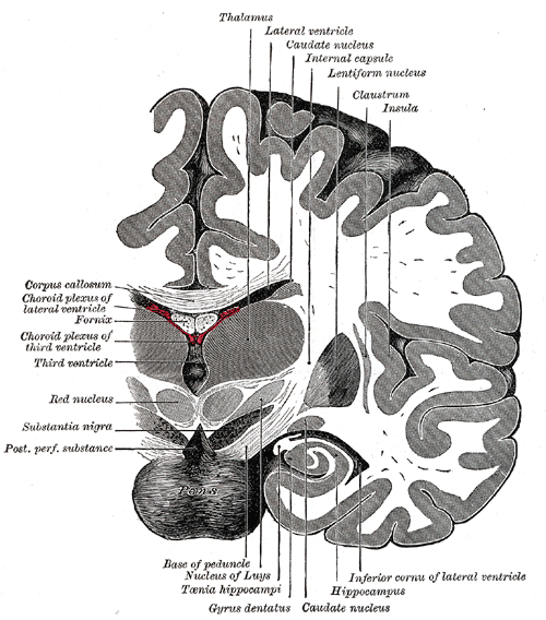
Uppermost part of the brainstem, around the cerebral aqueduct. It contains the CN III and IV nuclei, the substantia nigra and the corticospinal tracts in the cerebral peduncles, and is compressed in transtentorial herniation.
Core anatomy
- Location
Between the diencephalon above and the pons below, passing through the tentorial notch.
- Parts
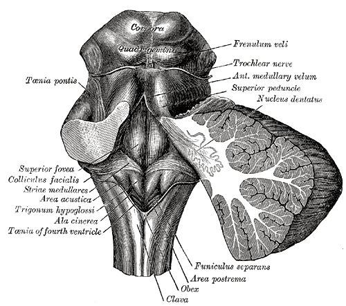
- Tectum (dorsal, behind the aqueduct): superior colliculi (visual reflexes, vertical gaze) and inferior colliculi (auditory pathway)
- Cerebral aqueduct (of Sylvius) connecting the third and fourth ventricles, surrounded by the periaqueductal gray (pain modulation)
- Tegmentum: nuclei of CN III (with Edinger–Westphal) and CN IV, the red nucleus, medial lemniscus, spinothalamic tract, MLF, reticular formation
- Substantia nigra (dopaminergic neurons of the pars compacta → striatum; see basal ganglia)
- Cerebral peduncles (crus cerebri): descending corticospinal and corticobulbar fibers; the interpeduncular fossa between them, where oculomotor nerve (CN III) emerges
- Pretectal area: pupillary light reflex relay (bilateral to both Edinger–Westphal nuclei → consensual response)
- Relations
The uncus of the temporal lobe and CN III lie against the free tentorial edge beside the peduncle; the pineal gland sits above the superior colliculi.
- Arterial supply
Branches of the posterior cerebral artery, superior cerebellar artery and top of the basilar artery (see vertebrobasilar system).
- Venous drainage
Basal veins → great cerebral vein (of Galen) → straight sinus.
- Notes
Contains the nuclei of CN III and IV; the trochlear nerve exits dorsally below the inferior colliculus (trochlear nerve (CN IV)).
Mentioned in
- Basal ganglia
- Corticospinal tract
- Internal capsule
- Oculomotor nerve (CN III)
- Pons
- Thalamus
- Trochlear nerve (CN IV)
- Vertebrobasilar system
Generated automatically from links in other entries.
Swipe sideways to see the whole diagram →


Clinical correlations
Parkinson disease PANCE
- Mechanism / lesion
Loss of dopaminergic neurons in the substantia nigra pars compacta (Lewy bodies, alpha-synuclein) → reduced dopamine in the striatum.
- Classic presentation
Resting (pill-rolling) tremor, rigidity (cogwheel), bradykinesia, postural instability; shuffling gait, masked face, micrographia; often asymmetric onset. Clinical diagnosis; levodopa-carbidopa.
Parinaud (dorsal midbrain) syndrome PANCE
- Mechanism / lesion
Compression of the dorsal midbrain/superior colliculi — pineal tumor (germinoma in young males), hydrocephalus, dorsal midbrain stroke.
- Classic presentation
Upward gaze palsy, light–near dissociation (pupils react to accommodation but not light), convergence–retraction nystagmus, eyelid retraction (Collier sign); often obstructive hydrocephalus from aqueduct compression.
Weber syndrome PANCE
- Mechanism / lesion
Infarct of the ventral (basal) midbrain — PCA/paramedian branches.
- Classic presentation
Ipsilateral CN III palsy + contralateral hemiparesis (cerebral peduncle).
Transtentorial (uncal) herniation and aqueductal stenosis PANCE
- Mechanism / lesion
A supratentorial mass pushes the uncus through the tentorial notch, compressing CN III, the peduncle and the PCA; congenital or acquired narrowing of the aqueduct blocks CSF flow.
- Classic presentation
Herniation: ipsilateral fixed dilated pupil, contralateral (or ipsilateral — Kernohan notch) hemiparesis, declining consciousness. Aqueductal stenosis: noncommunicating hydrocephalus with dilated lateral and third ventricles but a normal fourth ventricle.
High-yield PANCE pearls
- Substantia nigra degeneration → Parkinson disease (TRAP: tremor, rigidity, akinesia, postural instability).
- Pineal tumor → Parinaud syndrome (upgaze palsy, light–near dissociation) + hydrocephalus.
- Ipsilateral CN III + contralateral hemiparesis → Weber (midbrain).
- Dilated lateral and third ventricles, normal fourth → aqueductal stenosis.
Imaging anatomy
- CT
Noncontrast CT for herniation, hemorrhage and hydrocephalus.
- MRI
MRI is best for the brainstem (CT has bone artifact in the posterior fossa); DaT-SPECT can support a diagnosis of parkinsonism in unclear cases.
Physical exam
- Strength / function testing
Pupils (light and near response), vertical and horizontal eye movements, CN III/IV function; tone, tremor and bradykinesia (finger tapping), gait and pull test for parkinsonism.
Self-quiz
A 15-year-old boy has headaches, cannot look upward, and his pupils constrict when he looks at a near object but not to light. MRI shows a mass above the superior colliculi with dilated lateral and third ventricles. What is the diagnosis?
Correct answer: B
A pineal region tumor compressing the dorsal midbrain causes Parinaud syndrome (upgaze palsy, light–near dissociation, convergence–retraction nystagmus) and obstructive hydrocephalus at the aqueduct.
Show answer
B
A pineal region tumor compressing the dorsal midbrain causes Parinaud syndrome (upgaze palsy, light–near dissociation, convergence–retraction nystagmus) and obstructive hydrocephalus at the aqueduct.
A 66-year-old has a resting tremor of the right hand, cogwheel rigidity and slow finger tapping. Degeneration of which structure explains this?
Correct answer: B
Parkinson disease results from loss of dopaminergic neurons in the substantia nigra pars compacta. Caudate atrophy is Huntington disease; a subthalamic lesion causes hemiballismus.
Show answer
B
Parkinson disease results from loss of dopaminergic neurons in the substantia nigra pars compacta. Caudate atrophy is Huntington disease; a subthalamic lesion causes hemiballismus.
Weber syndrome (ipsilateral CN III palsy with contralateral hemiparesis) results from a lesion where?
Correct answer: B
Weber syndrome is a ventral (basal) midbrain infarct involving the CN III fascicles and the cerebral peduncle (corticospinal fibers). Dorsal midbrain lesions cause Parinaud syndrome.
Show answer
B
Weber syndrome is a ventral (basal) midbrain infarct involving the CN III fascicles and the cerebral peduncle (corticospinal fibers). Dorsal midbrain lesions cause Parinaud syndrome.
On MRI, which pattern of ventricular enlargement indicates aqueductal stenosis?
Correct answer: D
Obstruction at the cerebral aqueduct dilates everything upstream (lateral and third ventricles) while the fourth ventricle stays normal, a noncommunicating hydrocephalus.
Show answer
D
Obstruction at the cerebral aqueduct dilates everything upstream (lateral and third ventricles) while the fourth ventricle stays normal, a noncommunicating hydrocephalus.
Which midbrain area relays the pupillary light reflex to both Edinger–Westphal nuclei, producing the consensual response?
Correct answer: A
The pretectal nuclei receive optic tract input and project to both Edinger–Westphal nuclei, so light in one eye constricts both pupils (direct and consensual responses).
Show answer
A
The pretectal nuclei receive optic tract input and project to both Edinger–Westphal nuclei, so light in one eye constricts both pupils (direct and consensual responses).