Rectus abdominis
Long strap muscle on either side of the linea alba, enclosed in the rectus sheath. It flexes the trunk, and hematoma in its sheath is a classic complication of anticoagulation.
Core anatomy
- Location
Anterior abdominal wall, on each side of the linea alba, within the rectus sheath.
- Group
Anterolateral abdominal wall muscles (vertical group, with pyramidalis).
- Origin
Pubic symphysis and pubic crest.
- Insertion
Xiphoid process and the 5th–7th costal cartilages.
- Innervation
Thoracoabdominal nerves (anterior rami of T7–T11) and subcostal nerve (T12).
- Blood supply
Superior epigastric artery (from the internal thoracic artery) and inferior epigastric artery (from the external iliac artery), which anastomose within the sheath.
- Action
- Flexes the trunk (lumbar vertebral column)
- Compresses the abdominal viscera and increases intra-abdominal pressure
- Stabilizes the pelvis (e.g. during walking)
- Relations
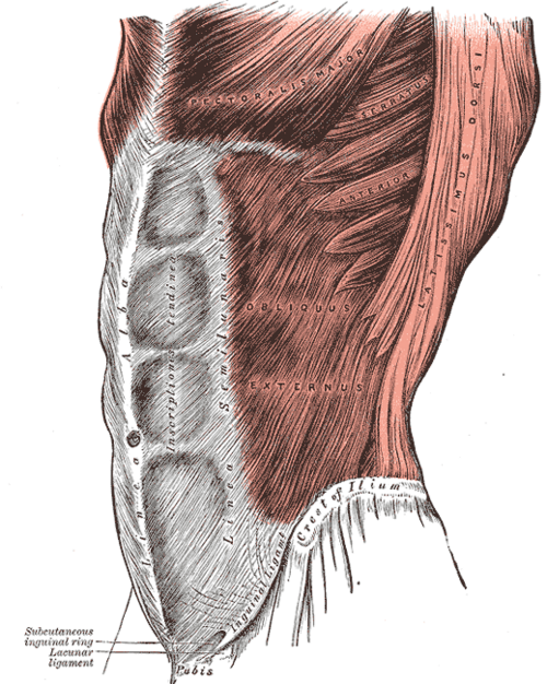
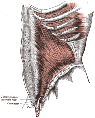
Usually three tendinous intersections attach it to the anterior layer of the sheath (the "six-pack"). Rectus sheath: above the arcuate line, the anterior layer is formed by the external oblique aponeurosis and the anterior lamina of the internal oblique aponeurosis, and the posterior layer by the posterior lamina of the internal oblique and the transversus abdominis aponeurosis. Below the arcuate line, all three aponeuroses pass anterior to the muscle, leaving only transversalis fascia behind it. The inferior epigastric vessels enter the sheath near the arcuate line.
Found in
Forms a boundary of
Mentioned in
Generated automatically from links in other entries.


Clinical correlations
Rectus sheath hematoma PANCE
- Mechanism / lesion
Rupture of the epigastric vessels or muscle fibers, typically in anticoagulated or older patients, often triggered by coughing, straining, trauma or injections. Below the arcuate line there is no posterior sheath, so the hematoma can spread and become large.
- Classic presentation
Acute abdominal pain with a tender, non-pulsatile abdominal wall mass that doesn't cross the midline and remains palpable when the muscles are tensed (Fothergill sign). Possible anemia.
Diastasis recti
- Mechanism / lesion
Widening of the linea alba between the two recti, common after pregnancy and in infants. It is not a true hernia (no fascial defect).
- Classic presentation
A midline ridge that bulges when the patient lifts the head from supine. Usually managed conservatively.
High-yield PANCE pearls
- Rectus sheath hematoma: anticoagulated patient + coughing → tender abdominal wall mass; confirm with CT or ultrasound.
- Below the arcuate line there is no posterior rectus sheath (only transversalis fascia).
- Diastasis recti is not a hernia; there is no fascial defect.
Imaging anatomy
- CT
CT is the test of choice for rectus sheath hematoma (a lenticular or spindle-shaped mass within the sheath; active extravasation on contrast).
- Ultrasound
Shows the muscle, sheath and hematoma. Measures inter-rectus distance in diastasis recti.
- Key views & landmarks
- CT abdomen with contrast (hematoma)
Physical exam
- Surface landmarks
The lateral border of the muscle forms the linea semilunaris, which crosses the costal margin at the tip of the 9th costal cartilage (the gallbladder fundus point on the right).
- Palpation
Palpate for a mass in the wall; ask the patient to tense the abdomen to see whether it stays palpable (wall) or disappears (intra-abdominal).
- Strength / function testing
Trunk flexion (sit-up) against resistance. Observe for midline bulging (diastasis) on head lift.
Self-quiz
A 74-year-old woman taking warfarin develops sudden left lower abdominal pain after a coughing fit. There is a tender mass in the lower abdominal wall that remains palpable when she tenses her abdominal muscles. What is the most likely diagnosis?
Correct answer: B
Rectus sheath hematoma is a classic complication in anticoagulated patients after coughing. A mass that remains palpable with the muscles tensed (Fothergill sign) lies in the wall, not the peritoneal cavity. Confirm with CT.
Show answer
B
Rectus sheath hematoma is a classic complication in anticoagulated patients after coughing. A mass that remains palpable with the muscles tensed (Fothergill sign) lies in the wall, not the peritoneal cavity. Confirm with CT.
Below the arcuate line, what forms the posterior boundary of rectus abdominis?
Correct answer: C
Below the arcuate line all three aponeuroses pass anterior to the muscle, so only transversalis fascia (and peritoneum) lies behind it.
Show answer
C
Below the arcuate line all three aponeuroses pass anterior to the muscle, so only transversalis fascia (and peritoneum) lies behind it.
Which nerves supply rectus abdominis?
Correct answer: B
Rectus abdominis is supplied by the anterior rami of T7–T11 (thoracoabdominal nerves) and T12 (subcostal nerve).
Show answer
B
Rectus abdominis is supplied by the anterior rami of T7–T11 (thoracoabdominal nerves) and T12 (subcostal nerve).