Inferior mesenteric artery
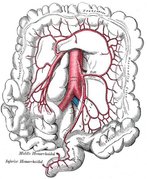
Artery of the hindgut, arising from the aorta at L3. It supplies the distal transverse colon to the upper rectum, and its ligation during aortic surgery puts the watershed colon at risk.
Core anatomy
- Location
Arises from the anterior aorta at about L3, behind the 3rd/4th part of the duodenum, 3–4 cm above the bifurcation.
- Origin
- Course
Descends retroperitoneally to the left, crossing the left common iliac artery to become the superior rectal artery in the pelvis.
- Branches
- Left colic artery (descending colon, distal transverse colon)
- Sigmoid arteries (sigmoid colon)
- Superior rectal artery (upper rectum), its terminal branch
- Supplies
Hindgut: distal third of the transverse colon, descending colon, sigmoid colon, upper rectum.
- Anastomoses
The marginal artery (of Drummond) along the colon connects it with the middle colic (superior mesenteric artery). The superior rectal artery anastomoses with the middle and inferior rectal arteries (internal iliac). Watershed zones: splenic flexure and rectosigmoid.
- Relations
The inferior mesenteric vein runs to its left and ascends to join the splenic vein. A horseshoe kidney is typically trapped beneath the IMA during its ascent.
Structures supplied
Mentioned in
Generated automatically from links in other entries.
Swipe sideways to see the whole diagram →

Clinical correlations
Ischemic colitis after aortic surgery PANCE
- Mechanism / lesion
The IMA is often sacrificed (or covered by an endograft) during AAA repair. If collaterals are poor, the left colon becomes ischemic.
- Classic presentation
Left-sided abdominal pain and bloody diarrhea after aortic repair (the sigmoid and splenic flexure are most affected).
Horseshoe kidney
- Mechanism / lesion
Fusion of the lower poles of the kidneys; ascent is halted when the isthmus catches under the IMA.
- Classic presentation
Usually asymptomatic. Increased risk of obstruction, stones and infection. Associated with Turner syndrome.
High-yield PANCE pearls
- IMA (L3) = hindgut: distal 1/3 transverse colon → upper rectum.
- Branches: left colic, sigmoid, superior rectal.
- Horseshoe kidney is trapped under the IMA.
- Post-AAA repair bloody diarrhea → ischemic colitis (IMA territory).
Imaging anatomy
- CT
CT angiography shows its origin, patency and the marginal artery; colonic wall thickening in ischemic colitis.
- Key views & landmarks
- CT angiography
- Colonoscopy/sigmoidoscopy to assess ischemic colitis after aortic surgery
Physical exam
- Strength / function testing
LLQ tenderness and bloody stool after aortic surgery should prompt evaluation for ischemic colitis.
Self-quiz
At approximately which vertebral level does the IMA arise?
Correct answer: D
The IMA arises at about L3; the celiac at T12 and the SMA at L1.
Show answer
D
The IMA arises at about L3; the celiac at T12 and the SMA at L1.
A horseshoe kidney fails to ascend normally because the isthmus becomes trapped under which structure?
Correct answer: C
The fused lower poles of a horseshoe kidney catch on the inferior mesenteric artery during ascent.
Show answer
C
The fused lower poles of a horseshoe kidney catch on the inferior mesenteric artery during ascent.
Which is the terminal branch of the IMA?
Correct answer: C
The IMA continues as the superior rectal artery into the pelvis. The middle rectal artery comes from the internal iliac; the middle colic from the SMA.
Show answer
C
The IMA continues as the superior rectal artery into the pelvis. The middle rectal artery comes from the internal iliac; the middle colic from the SMA.
Which artery is NOT a branch of the inferior mesenteric artery?
Correct answer: B
The IMA gives the left colic, sigmoid and superior rectal arteries. The middle colic artery is a branch of the SMA; the marginal artery connects the two territories.
Show answer
B
The IMA gives the left colic, sigmoid and superior rectal arteries. The middle colic artery is a branch of the SMA; the marginal artery connects the two territories.
The IMA supplies which part of the gut?
Correct answer: D
The IMA (L3) supplies the hindgut: distal third of the transverse colon, descending and sigmoid colon, and upper rectum. The SMA supplies the midgut; the anal canal below the pectinate line is supplied by the inferior rectal arteries.
Show answer
D
The IMA (L3) supplies the hindgut: distal third of the transverse colon, descending and sigmoid colon, and upper rectum. The SMA supplies the midgut; the anal canal below the pectinate line is supplied by the inferior rectal arteries.
Flashcards
See also
- Colon
- Abdominal aorta
- Superior mesenteric artery
- Kidney
- Foregut, midgut and hindgut (comparison table)
- Anal canal above vs below the pectinate line (comparison table)