Muscles of facial expression
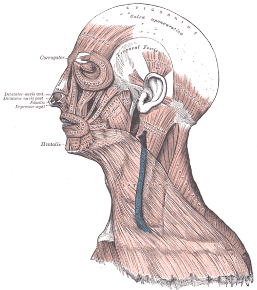
Thin subcutaneous muscles derived from the second pharyngeal arch and supplied by the facial nerve (CN VII). Their pattern of weakness distinguishes a peripheral facial palsy (Bell palsy) from a stroke.
Core anatomy
- Location
In the superficial fascia of the face, scalp and neck; most insert into the skin.
- Group
Second pharyngeal arch derivatives
- Origin
Mostly from bones of the face or fascia (e.g. orbicularis oculi from the medial orbital margin; zygomaticus major from the zygomatic bone; buccinator from the alveolar processes and pterygomandibular raphe).
- Insertion
Mostly into the skin and each other (e.g. modiolus at the angle of the mouth), which is why they move the skin.
- Innervation
Facial nerve (CN VII) — its five terminal branches leave the parotid gland: temporal, zygomatic, buccal, marginal mandibular and cervical ("To Zanzibar By Motor Car"). Also stapedius, stylohyoid and the posterior belly of digastric.
- Blood supply
Facial, superficial temporal and transverse facial arteries (branches of the external carotid artery).
- Action
- Orbicularis oculi: closes the eyelids (blinking, tight closure)
- Frontalis (occipitofrontalis): raises the eyebrows, wrinkles the forehead
- Orbicularis oris: closes and purses the lips
- Buccinator: compresses the cheek (keeps food between the teeth; blowing)
- Zygomaticus major: smiling; depressor anguli oris: frowning
- Platysma: tenses the skin of the neck, depresses the mandible
- Relations
The upper face (frontalis, upper orbicularis oculi) receives bilateral cortical input, whereas the lower face receives mainly contralateral input — the key to localizing facial weakness.
Swipe sideways to see the whole diagram →

Clinical correlations
Bell palsy (peripheral facial nerve palsy) PANCE
- Mechanism / lesion
Idiopathic (often attributed to HSV reactivation) swelling of the facial nerve in the facial canal — a lower motor neuron lesion.
- Classic presentation
Sudden unilateral weakness of the whole face, including the forehead (can't raise the eyebrow or close the eye), drooping mouth, loss of nasolabial fold; may have hyperacusis, loss of taste to the anterior two-thirds of the tongue, and decreased tearing. Treat with oral corticosteroids within 72 hours (± antivirals) and eye protection.
Central (upper motor neuron) facial palsy PANCE
- Mechanism / lesion
Stroke or tumor affecting the corticobulbar fibers above the facial nucleus.
- Classic presentation
Weakness of the lower face only on the opposite side, with the forehead spared (bilateral upper-face innervation), usually with other deficits (arm weakness, aphasia).
Other causes of peripheral facial palsy PANCE
- Mechanism / lesion
Ramsay Hunt syndrome (VZV — vesicles in the ear canal), Lyme disease (may be bilateral), parotid tumors or surgery, temporal bone fracture, otitis media, sarcoidosis.
- Classic presentation
Look in the ear for vesicles, ask about tick exposure, and palpate the parotid; bilateral palsy suggests Lyme, Guillain–Barré or sarcoidosis.
High-yield PANCE pearls
- Forehead involved = peripheral (Bell); forehead spared = central (stroke).
- Bell palsy → prednisone within 72 h + eye care (lubricant, taping).
- Vesicles in the ear + facial palsy → Ramsay Hunt (VZV).
- Bilateral facial palsy → Lyme, Guillain–Barré, sarcoidosis.
- Facial nerve branches: Temporal, Zygomatic, Buccal, Marginal mandibular, Cervical.
Imaging anatomy
- CT
Head CT if stroke is suspected (forehead-sparing weakness, other deficits); temporal bone CT for trauma.
- MRI
MRI with contrast for atypical or progressive palsy, recurrence, or no recovery after about 3 months (tumor along the nerve, e.g. parotid or cerebellopontine angle).
Physical exam
- Surface landmarks
The facial nerve branches fan out from the anterior border of the parotid gland.
- Strength / function testing
Ask the patient to raise the eyebrows (frontalis), close the eyes tightly against resistance (orbicularis oculi), smile and show the teeth, puff the cheeks (buccinator), and tense the neck (platysma). Look for Bell phenomenon (the eye rolls up when trying to close). Check taste, hearing (hyperacusis) and the ear canal.
Self-quiz
A 35-year-old woman wakes with drooping of the right side of her face. She cannot raise her right eyebrow or fully close her right eye. There are no other neurologic deficits and the ear canal is normal. What is the best treatment?
Correct answer: B
Involvement of the forehead indicates a peripheral (lower motor neuron) lesion — Bell palsy. Corticosteroids within 72 hours improve recovery; protect the eye from exposure keratitis. tPA is for ischemic stroke, which spares the forehead.
Show answer
B
Involvement of the forehead indicates a peripheral (lower motor neuron) lesion — Bell palsy. Corticosteroids within 72 hours improve recovery; protect the eye from exposure keratitis. tPA is for ischemic stroke, which spares the forehead.
Why is the forehead spared in a stroke causing facial weakness?
Correct answer: B
The part of the facial nucleus supplying the upper face receives input from both cerebral hemispheres, so a unilateral cortical lesion spares the forehead. The lower face receives mainly contralateral input.
Show answer
B
The part of the facial nucleus supplying the upper face receives input from both cerebral hemispheres, so a unilateral cortical lesion spares the forehead. The lower face receives mainly contralateral input.
A 60-year-old has painful vesicles in the right ear canal and new weakness of the whole right side of the face. What is the diagnosis?
Correct answer: E
Ramsay Hunt syndrome is reactivation of varicella-zoster in the geniculate ganglion, causing facial palsy with vesicles in the ear canal. Always look in the ear in a facial palsy.
Show answer
E
Ramsay Hunt syndrome is reactivation of varicella-zoster in the geniculate ganglion, causing facial palsy with vesicles in the ear canal. Always look in the ear in a facial palsy.
Bilateral peripheral facial palsy should raise suspicion for which conditions?
Correct answer: A
Bell palsy is almost always unilateral. Bilateral facial palsy suggests Lyme disease, Guillain–Barré syndrome or sarcoidosis.
Show answer
A
Bell palsy is almost always unilateral. Bilateral facial palsy suggests Lyme disease, Guillain–Barré syndrome or sarcoidosis.
Which nerve supplies the buccinator muscle?
Correct answer: D
Buccinator is a muscle of facial expression (2nd arch), supplied by the buccal branch of the facial nerve. The long buccal nerve of V3 is purely sensory to the cheek.
Show answer
D
Buccinator is a muscle of facial expression (2nd arch), supplied by the buccal branch of the facial nerve. The long buccal nerve of V3 is purely sensory to the cheek.