Celiac trunk
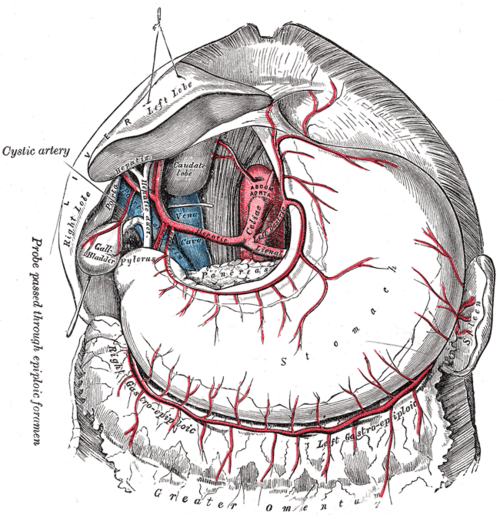
Short anterior branch of the abdominal aorta at T12 and the artery of the foregut. It divides into the left gastric, splenic and common hepatic arteries.
Core anatomy
- Location
Arises from the anterior aorta just below the aortic hiatus, at about T12, above the pancreas.
- Origin
- Course
A short trunk (1–2 cm) surrounded by the celiac plexus and nodes; it divides almost immediately.
- Branches
- Left gastric artery: along the lesser curvature of the stomach; esophageal branches
- Splenic artery: tortuous course along the superior border of the pancreas to the spleen; gives pancreatic branches, short gastric, left gastro-omental and posterior gastric arteries
- Common hepatic artery → gastroduodenal artery (→ right gastro-omental, superior pancreaticoduodenal) and hepatic artery proper (→ right gastric, right and left hepatic arteries; the cystic artery usually comes from the right hepatic)
- Supplies
Foregut derivatives: abdominal esophagus, stomach, duodenum proximal to the major papilla, liver, gallbladder, pancreas (with the SMA), and the spleen (a mesodermal organ in the foregut mesentery).
- Anastomoses
Pancreaticoduodenal arcades connect it with the superior mesenteric artery, an important collateral route in occlusive disease.
Structures supplied
Mentioned in
Generated automatically from links in other entries.
Swipe sideways to see the whole diagram →

Clinical correlations
Median arcuate ligament syndrome
- Mechanism / lesion
Compression of the celiac trunk by the median arcuate ligament of the diaphragm (worse in expiration).
- Classic presentation
Postprandial epigastric pain, weight loss, and an epigastric bruit that varies with respiration, typically in young thin women. A diagnosis of exclusion.
Upper GI bleeding from celiac branches PANCE
- Mechanism / lesion
Ulcers eroding branches: posterior duodenal ulcer → gastroduodenal artery; lesser-curvature gastric ulcer → left gastric artery; posterior gastric ulcer → splenic artery.
- Classic presentation
Hematemesis or melena, hemodynamic instability. Managed endoscopically or with angiographic embolization.
Splenic artery aneurysm
- Mechanism / lesion
The most common visceral artery aneurysm; more common in women, and associated with pregnancy and portal hypertension.
- Classic presentation
Usually asymptomatic. Rupture in pregnancy carries high maternal and fetal mortality.
High-yield PANCE pearls
- Celiac trunk (T12) → left gastric, splenic, common hepatic.
- Foregut = esophagus (abdominal) to the proximal duodenum, liver, gallbladder, pancreas (+ spleen).
- Posterior duodenal ulcer bleeds from the gastroduodenal artery.
Imaging anatomy
- CT
CT angiography shows celiac stenosis (a characteristic hooked narrowing in median arcuate ligament syndrome), aneurysms and variant anatomy.
- Ultrasound
Duplex shows elevated celiac velocities in stenosis; in median arcuate ligament syndrome velocities increase in expiration.
- Key views & landmarks
- CT angiography (sagittal view of the celiac and SMA origins)
- Catheter angiography for embolization of bleeding branches
Physical exam
- Strength / function testing
Listen for an epigastric bruit (celiac or SMA stenosis). Localize foregut pain to the epigastrium.
Self-quiz
Which of the following is NOT a direct branch of the celiac trunk?
Correct answer: D
The celiac trunk gives the left gastric, splenic and common hepatic arteries. The gastroduodenal artery comes from the common hepatic artery.
Show answer
D
The celiac trunk gives the left gastric, splenic and common hepatic arteries. The gastroduodenal artery comes from the common hepatic artery.
At approximately what vertebral level does the celiac trunk arise?
Correct answer: C
The celiac trunk arises at T12, the SMA at L1, and the IMA at L3.
Show answer
C
The celiac trunk arises at T12, the SMA at L1, and the IMA at L3.
The short gastric arteries arise from which vessel?
Correct answer: B
The short gastric arteries (supplying the fundus) and the left gastro-omental artery are branches of the splenic artery.
Show answer
B
The short gastric arteries (supplying the fundus) and the left gastro-omental artery are branches of the splenic artery.
A thin 28-year-old woman has postprandial epigastric pain and weight loss. There is an epigastric bruit that changes with respiration, and other causes have been excluded. What is the most likely diagnosis?
Correct answer: D
Median arcuate ligament syndrome is compression of the celiac trunk by the median arcuate ligament of the diaphragm (worse in expiration), typically in young thin women. It is a diagnosis of exclusion.
Show answer
D
Median arcuate ligament syndrome is compression of the celiac trunk by the median arcuate ligament of the diaphragm (worse in expiration), typically in young thin women. It is a diagnosis of exclusion.
What is the most common visceral artery aneurysm, notable for its risk of rupture in pregnancy?
Correct answer: B
The splenic artery aneurysm is the most common visceral artery aneurysm, more common in women and associated with pregnancy and portal hypertension. Rupture in pregnancy carries high maternal and fetal mortality.
Show answer
B
The splenic artery aneurysm is the most common visceral artery aneurysm, more common in women and associated with pregnancy and portal hypertension. Rupture in pregnancy carries high maternal and fetal mortality.