Atlas and axis (C1 and C2)
The first two cervical vertebrae, specialized for head movement. The atlas (C1) has no body; the axis (C2) carries the dens, around which the atlas rotates. Their fractures and instability are high-yield.
Core anatomy
- Location
Upper neck, between the occiput and C3.
- Classification
Atypical cervical vertebrae.
- Key features & landmarks
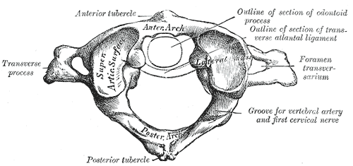
- Atlas (C1): ring of anterior and posterior arches with two lateral masses; no body and no spinous process. Superior articular facets receive the occipital condyles; a groove on the posterior arch carries the vertebral artery
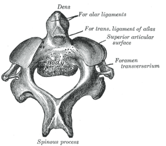
- Axis (C2): strong body projecting upward as the dens (odontoid process), the embryologic body of C1. Large spinous process (first palpable below the occiput)
- Transverse ligament of the atlas holds the dens against the anterior arch; alar and apical ligaments tie the dens to the skull; the tectorial membrane continues the posterior longitudinal ligament
- Relations
Atlanto-occipital joints: flexion and extension (nodding "yes"). Atlantoaxial joints: rotation (shaking "no"); they provide roughly half of cervical rotation. The spinal cord and medulla lie just behind the dens.


Clinical correlations
Jefferson fracture (C1 burst) PANCE
- Mechanism / lesion
Axial loading onto the top of the head (e.g. diving into shallow water) drives the occipital condyles into the lateral masses, breaking the ring in several places.
- Classic presentation
Upper neck pain, often without neurologic deficit (the canal widens). On the open-mouth view, the lateral masses overhang C2. Stability depends on the transverse ligament.
Odontoid (dens) fractures PANCE
- Mechanism / lesion
Falls in older adults (hyperextension or flexion) or high-energy trauma in younger patients.
- Classic presentation
Upper neck pain, sometimes minimal findings in older adults. Classified by level; type II (base of the dens) is the most common and has a high risk of nonunion (poor blood supply).
Hangman fracture PANCE
- Mechanism / lesion
Bilateral fractures of the pars interarticularis/pedicles of C2 from hyperextension with distraction (judicial hanging historically; now usually vehicle collisions).
- Classic presentation
Neck pain; traumatic spondylolisthesis of C2 on C3. Neurologic injury is less common because the canal widens.
Atlantoaxial instability PANCE
- Mechanism / lesion
Laxity or erosion of the transverse ligament, as in Down syndrome and rheumatoid arthritis (pannus).
- Classic presentation
Neck pain, occipital headache, or myelopathy. Risk of cord injury during neck manipulation or intubation. Patients with long-standing RA may need flexion–extension films before surgery. An increased atlantodental interval on lateral films suggests instability. verify
High-yield PANCE pearls
- Jefferson = C1 burst from axial load (diving).
- Hangman = C2 pars/pedicle fractures from hyperextension.
- Odontoid type II fracture is most common and prone to nonunion.
- RA and Down syndrome → atlantoaxial instability (transverse ligament); caution with intubation.
- Atlanto-occipital = "yes" (nodding); atlantoaxial = "no" (rotation).
Imaging anatomy
- X-ray
Open-mouth (odontoid) AP view shows the dens and lateral masses of C1 (overhang in Jefferson fracture). Lateral view: atlantodental interval, prevertebral swelling. Flexion–extension views for instability (only in stable, alert patients).
- CT
CT is the test of choice for suspected C1–C2 fractures.
- MRI
Transverse ligament integrity, cord compression, soft-tissue injury.
- Key views & landmarks
- Open-mouth odontoid view
- CT cervical spine with sagittal and coronal reformats
Physical exam
- Surface landmarks
C2 spinous process is the first bony prominence palpable in the midline below the external occipital protuberance. The transverse process of C1 can be felt between the mastoid process and the angle of the mandible.
- Strength / function testing
Neck rotation (mostly atlantoaxial) and nodding (atlanto-occipital) — only once fracture is excluded in trauma. Full neurologic exam for myelopathy.
Self-quiz
A 19-year-old dives into a shallow pool and has upper neck pain. The open-mouth view shows the lateral masses of C1 displaced beyond the edges of C2. What is the injury?
Correct answer: B
Axial load from diving causes a Jefferson (C1 burst) fracture; lateral mass overhang on the open-mouth view is characteristic. A hangman fracture involves the C2 pars from hyperextension.
Show answer
B
Axial load from diving causes a Jefferson (C1 burst) fracture; lateral mass overhang on the open-mouth view is characteristic. A hangman fracture involves the C2 pars from hyperextension.
Which patient is at greatest risk of atlantoaxial subluxation during intubation?
Correct answer: A
Rheumatoid arthritis pannus can erode the transverse ligament, causing atlantoaxial instability. Down syndrome is another classic association.
Show answer
A
Rheumatoid arthritis pannus can erode the transverse ligament, causing atlantoaxial instability. Down syndrome is another classic association.
Which movement occurs mainly at the atlantoaxial joints?
Correct answer: B
The atlantoaxial joints provide about half of cervical rotation; the atlanto-occipital joints allow nodding.
Show answer
B
The atlantoaxial joints provide about half of cervical rotation; the atlanto-occipital joints allow nodding.
After a head-on collision with neck hyperextension, CT shows bilateral fractures through the pars interarticularis of C2 with anterior slip of C2 on C3. What is this injury called?
Correct answer: C
A hangman fracture is a bilateral fracture of the C2 pars interarticularis/pedicles from hyperextension with distraction, producing traumatic spondylolisthesis of C2 on C3. The canal widens, so neurologic injury is less common. A Jefferson fracture is a C1 burst from axial load.
Show answer
C
A hangman fracture is a bilateral fracture of the C2 pars interarticularis/pedicles from hyperextension with distraction, producing traumatic spondylolisthesis of C2 on C3. The canal widens, so neurologic injury is less common. A Jefferson fracture is a C1 burst from axial load.
Which odontoid (dens) fracture is most common and most prone to nonunion?
Correct answer: B
Type II fractures through the base of the dens are the most common and have a high risk of nonunion because of poor blood supply. They are common after falls in older adults.
Show answer
B
Type II fractures through the base of the dens are the most common and have a high risk of nonunion because of poor blood supply. They are common after falls in older adults.