Trigeminal nerve (CN V)
Largest cranial nerve — sensation from the face, scalp, eye, nose and mouth in three divisions, plus motor supply to the muscles of mastication. Trigeminal neuralgia and shingles of V1 are the classic problems.
Core anatomy
- Origin
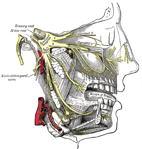
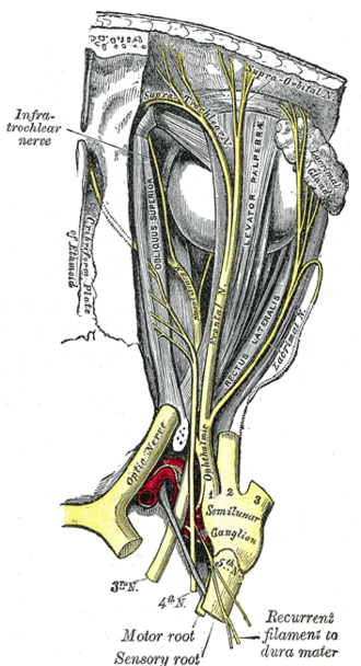
Leaves the lateral pons as a large sensory root and a small motor root. Nuclei: principal sensory (pons — touch), spinal nucleus (down into the cervical cord — pain and temperature), mesencephalic (proprioception, jaw jerk) and motor nuclei. The sensory cell bodies form the trigeminal (semilunar) ganglion in Meckel cave on the petrous apex.
- Course
The ganglion gives three divisions: V1 ophthalmic — lateral wall of the cavernous sinus → superior orbital fissure; V2 maxillary — lateral wall of the cavernous sinus → foramen rotundum → pterygopalatine fossa; V3 mandibular (with the whole motor root) → foramen ovale → infratemporal fossa. Mnemonic "Standing Room Only" (SOF, rotundum, ovale).
- Branches
- V1 ophthalmic (sensory): frontal (supraorbital, supratrochlear), lacrimal, nasociliary (cornea, tip of the nose) — forehead, upper eyelid, cornea, dorsum of nose
- V2 maxillary (sensory): infraorbital, zygomatic, superior alveolar, palatine and nasal branches — cheek, lower eyelid, upper lip, upper teeth, palate, nasal cavity
- V3 mandibular (sensory + motor): auriculotemporal, buccal, lingual (anterior 2/3 of tongue), inferior alveolar (lower teeth, chin via the mental nerve) and motor branches
- Motor
V3 only: Muscles of mastication (masseter, temporalis, medial and lateral pterygoids), mylohyoid, anterior belly of digastric, tensor tympani, tensor veli palatini.
- Sensory
Face and anterior scalp to the vertex, cornea and conjunctiva, nasal and oral cavities, teeth, anterior two-thirds of the tongue (general sensation), most of the supratentorial dura. The angle of the jaw is supplied by C2–C3 (great auricular nerve), not CN V.
- Relations
V1 and V2 run in the lateral wall of the cavernous sinus with CN III and IV (see dural venous sinuses). Parasympathetic fibers from CN VII and IX "hitchhike" on V branches to the lacrimal, nasal, palatine and parotid glands.
Muscles innervated
- Muscles of mastication — Mandibular division of the trigeminal nerve (V3) — motor root
Other structures innervated
Found in
Mentioned in
Generated automatically from links in other entries.
Swipe sideways to see the whole diagram →


Clinical correlations
Trigeminal neuralgia PANCE
- Mechanism / lesion
Usually vascular compression of the root near the pons (superior cerebellar artery); in a young patient or with bilateral symptoms, think multiple sclerosis.
- Classic presentation
Brief (seconds), electric, stabbing unilateral facial pain, usually V2 or V3, triggered by chewing, talking, brushing teeth, touching the face or wind; normal neurologic exam. Carbamazepine (or oxcarbazepine) is first-line; microvascular decompression or ablation if refractory. MRI to exclude secondary causes.
Herpes zoster ophthalmicus PANCE
- Mechanism / lesion
Reactivation of varicella-zoster virus in the trigeminal ganglion, V1 distribution.
- Classic presentation
Painful vesicular rash on the forehead and upper eyelid; Hutchinson sign (vesicles on the tip of the nose — nasociliary branch) predicts eye involvement (keratitis, uveitis). Oral antivirals within 72 hours and urgent ophthalmology.
Motor (V3) lesion
- Mechanism / lesion
Tumors at the skull base or foramen ovale, trauma.
- Classic presentation
Weak jaw clench and masseter/temporalis wasting; the jaw deviates toward the weak side on opening (unopposed contralateral lateral pterygoid).
High-yield PANCE pearls
- Lightning-like facial pain triggered by chewing/touch → trigeminal neuralgia → carbamazepine.
- Trigeminal neuralgia in a young adult → consider MS.
- Zoster with vesicles on the nose tip (Hutchinson sign) → eye involvement → ophthalmology.
- Corneal reflex: afferent V1, efferent VII.
- Jaw deviates toward a V3 lesion.
- SOF (V1), rotundum (V2), ovale (V3) — "Standing Room Only".
Imaging anatomy
- CT
CT of the skull base foramina for tumors or fractures.
- MRI
MRI brain with thin-section imaging of the trigeminal root (e.g. FIESTA/CISS) to show vascular compression or exclude tumor or MS plaques.
Physical exam
- Surface landmarks
Supraorbital (V1), infraorbital (V2) and mental (V3) foramina lie on a vertical line through the pupil.
- Palpation
Palpate masseter and temporalis while the patient clenches the teeth.
- Strength / function testing
Light touch and pinprick (± temperature) in each of the three divisions on both sides (forehead, cheek, jaw — not the angle of the jaw). Motor: clench teeth (palpate masseter/temporalis), open the jaw against resistance (deviation). Jaw jerk (brisk in upper motor neuron lesions above the pons).
Corneal reflex
- Technique
Touch the cornea (not the conjunctiva) from the side with a wisp of cotton while the patient looks away.
- Positive test
Absent blink in both eyes when one cornea is touched.
- Significance
Afferent V1 (nasociliary), efferent CN VII (orbicularis oculi). Loss with preserved blink on stimulating the other eye → V1 lesion (e.g. acoustic neuroma compressing V).
Self-quiz
A 62-year-old woman has attacks of stabbing pain in the right cheek lasting a few seconds, set off by brushing her teeth. Neurologic exam is normal. What is the first-line treatment?
Correct answer: B
Brief, triggered, electric pain in V2 with a normal exam is trigeminal neuralgia; carbamazepine is first-line. Obtain MRI to exclude a secondary cause.
Show answer
B
Brief, triggered, electric pain in V2 with a normal exam is trigeminal neuralgia; carbamazepine is first-line. Obtain MRI to exclude a secondary cause.
The motor fibers of the trigeminal nerve exit the skull through which foramen?
Correct answer: C
All motor fibers travel with V3 (mandibular), which leaves through the foramen ovale. The foramen spinosum transmits the middle meningeal artery.
Show answer
C
All motor fibers travel with V3 (mandibular), which leaves through the foramen ovale. The foramen spinosum transmits the middle meningeal artery.
A patient has a vesicular rash over the left forehead and upper eyelid, with vesicles on the tip of the nose. What does the nasal involvement indicate?
Correct answer: B
Hutchinson sign — the nasociliary branch of V1 supplies both the nose tip and the eye, so nasal vesicles predict ocular zoster. Urgent ophthalmology and antivirals.
Show answer
B
Hutchinson sign — the nasociliary branch of V1 supplies both the nose tip and the eye, so nasal vesicles predict ocular zoster. Urgent ophthalmology and antivirals.
Which nerve supplies the skin over the angle of the jaw?
Correct answer: B
The angle of the jaw is supplied by the great auricular nerve (C2–C3), not CN V, which helps tell trigeminal from cervical sensory loss.
Show answer
B
The angle of the jaw is supplied by the great auricular nerve (C2–C3), not CN V, which helps tell trigeminal from cervical sensory loss.
A 28-year-old develops trigeminal neuralgia. Which underlying condition should be considered?
Correct answer: E
Trigeminal neuralgia is usually from vascular compression in older adults. In a young patient or with bilateral symptoms, consider multiple sclerosis and obtain MRI.
Show answer
E
Trigeminal neuralgia is usually from vascular compression in older adults. In a young patient or with bilateral symptoms, consider multiple sclerosis and obtain MRI.