Urinary bladder
Distensible muscular reservoir for urine lying behind the pubis. Its autonomic control explains neurogenic bladder, and rupture type (intraperitoneal vs extraperitoneal) decides management.
Core anatomy
- Location
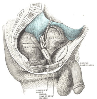
When empty, in the lesser pelvis behind the pubic symphysis, separated from it by the retropubic (prevesical) space. As it fills it rises extraperitoneally into the lower abdomen, lifting the peritoneum off the anterior abdominal wall.
- Parts
- Apex: points toward the symphysis; continues as the median umbilical ligament (obliterated urachus) to the umbilicus
- Body and fundus (base): the base faces posteroinferiorly toward the rectum (male) or vagina (female)
- Neck: where the base and inferolateral surfaces meet, around the internal urethral orifice; rests on the prostate in males
- Trigone: smooth triangle between the two ureteric orifices and the internal urethral orifice (derived from the mesonephric ducts, so it lacks rugae)
- Detrusor muscle: interlacing smooth muscle forming the wall; in males a circular internal urethral sphincter at the neck prevents retrograde ejaculation
- Relations
Superior surface covered by peritoneum; in females the uterus lies on it (vesicouterine pouch). Males: the ductus deferens and seminal glands, seminal glands and rectum lie posterior to the base. The ureters enter the base obliquely, which forms a flap-valve against reflux.
- Arterial supply
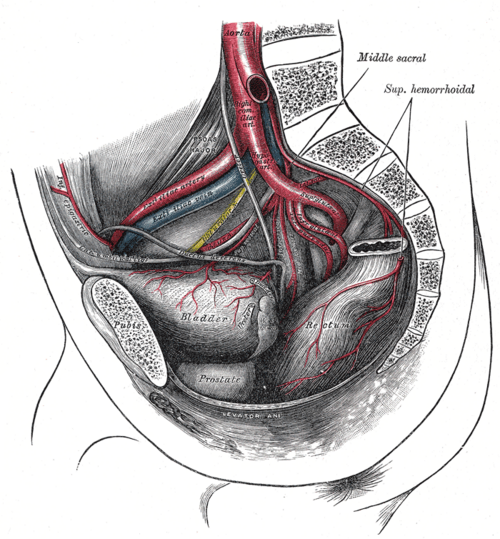
Superior vesical arteries (from the patent part of the umbilical artery) and inferior vesical arteries (males) or vaginal arteries (females), all from the anterior division of the internal iliac artery.
- Venous drainage
Vesical venous plexus (continuous with the prostatic plexus in males) → internal iliac veins.
- Lymphatic drainage
External and internal iliac nodes (see pelvic and inguinal lymph drainage).
- Innervation
Parasympathetic (pelvic splanchnic nerves, S2–S4; muscarinic M3) → detrusor contraction (micturition). Sympathetic (T11–L2 via the hypogastric plexuses) → detrusor relaxation (β3) and contraction of the bladder neck/internal sphincter (α1) = storage. Somatic: Pudendal nerve (S2–S4) → voluntary external urethral sphincter. See pelvic autonomic nerves.
Mentioned in
Generated automatically from links in other entries.


Clinical correlations
Bladder rupture PANCE
- Mechanism / lesion
Extraperitoneal (most common): associated with pelvic fractures (bone spicules or shearing). Intraperitoneal: a blow to the lower abdomen when the bladder is full bursts the dome, the weakest part covered by peritoneum.
- Classic presentation
Gross hematuria, suprapubic pain, inability to void. Intraperitoneal rupture causes urinary ascites, peritonitis and a rising creatinine (peritoneal reabsorption).
- Notes
Extraperitoneal rupture is usually managed with catheter drainage; intraperitoneal rupture needs surgical repair.
Acute urinary retention PANCE
- Mechanism / lesion
Outlet obstruction (BPH most common in men), anticholinergic or opioid drugs, constipation, postoperative state, or neurologic causes (spinal cord injury, cauda equina syndrome, diabetic neuropathy).
- Classic presentation
Painful inability to void with a palpable, dull suprapubic mass; overflow incontinence in chronic retention. Treat with catheter drainage and address the cause.
Urge incontinence (overactive bladder) PANCE
- Mechanism / lesion
Involuntary detrusor contractions (detrusor overactivity).
- Classic presentation
Sudden strong urge followed by leakage, frequency and nocturia. First-line: behavioral therapy (bladder training, fluid and caffeine reduction). Then antimuscarinics (e.g. oxybutynin; anticholinergic side effects, caution in older adults) or the β3-agonist mirabegron.
Bladder cancer PANCE
- Mechanism / lesion
Usually urothelial carcinoma. Risk factors — smoking (most important), aromatic amines/aniline dyes, cyclophosphamide; Schistosoma haematobium causes squamous cell carcinoma.
- Classic presentation
Painless gross hematuria in an older smoker. Evaluate with cystoscopy plus CT urography.
Urachal anomalies
- Mechanism / lesion
Failure of the urachus (allantois remnant) to obliterate.
- Classic presentation
Patent urachus leaks urine from the umbilicus; urachal cyst or sinus may become infected; urachal adenocarcinoma arises at the bladder dome.
High-yield PANCE pearls
- Pelvic fracture + hematuria → extraperitoneal rupture (catheter). Full bladder + blow → intraperitoneal rupture at the dome (surgery).
- Parasympathetic S2–S4 = pee (detrusor contraction); sympathetic = storage.
- Urge incontinence → behavioral therapy, then oxybutynin or mirabegron.
- Painless gross hematuria in a smoker = bladder cancer until proven otherwise → cystoscopy.
- Urine draining from the umbilicus in a newborn → patent urachus.
Imaging anatomy
- X-ray
Retrograde cystography (plain film) is an older method for detecting rupture.
- CT
CT cystography (retrograde filling of the bladder with dilute contrast through a catheter) is the test of choice for suspected rupture: contrast outlining bowel loops = intraperitoneal; "flame-shaped" or "molar tooth" extravasation around the bladder = extraperitoneal. CT urography for hematuria evaluation of the upper tracts.
- Ultrasound
Bedside bladder scan measures post-void residual (PVR) volume. Ultrasound shows wall thickening, masses, stones, and ureteral jets.
- Key views & landmarks
- CT cystogram (bladder distended with contrast)
- Voiding cystourethrogram (VCUG) for vesicoureteral reflux in children
Physical exam
- Surface landmarks
The empty bladder lies behind the symphysis and can't be felt; a distended bladder rises toward the umbilicus.
- Palpation
A distended bladder is a smooth, midline, dull-to-percussion suprapubic mass that may cause an urge to void when pressed.
- Strength / function testing
Measure PVR after voiding (bladder scan or catheter). Neurologic exam for neurogenic bladder: saddle sensation, anal tone, bulbocavernosus reflex (S2–S4). Suprapubic catheters are inserted in the midline about 2 finger-breadths above the symphysis into a distended bladder, staying extraperitoneal.
Self-quiz
After a night of drinking, a 27-year-old man is punched in the lower abdomen. He has diffuse abdominal pain, gross hematuria and a rising serum creatinine. CT cystography shows contrast outlining loops of bowel. What is the most appropriate management?
Correct answer: B
A blow to a full bladder ruptures the dome into the peritoneal cavity (contrast around bowel loops). Intraperitoneal rupture requires surgical repair; urine absorbed across the peritoneum raises creatinine. Extraperitoneal ruptures are usually managed with catheter drainage.
Show answer
B
A blow to a full bladder ruptures the dome into the peritoneal cavity (contrast around bowel loops). Intraperitoneal rupture requires surgical repair; urine absorbed across the peritoneum raises creatinine. Extraperitoneal ruptures are usually managed with catheter drainage.
Which receptor mediates detrusor contraction during micturition?
Correct answer: C
Parasympathetic pelvic splanchnic nerves (S2–S4) release acetylcholine onto M3 receptors on the detrusor, which is why antimuscarinics treat overactive bladder. β3 stimulation relaxes the detrusor (mirabegron); α1 contracts the bladder neck.
Show answer
C
Parasympathetic pelvic splanchnic nerves (S2–S4) release acetylcholine onto M3 receptors on the detrusor, which is why antimuscarinics treat overactive bladder. β3 stimulation relaxes the detrusor (mirabegron); α1 contracts the bladder neck.
A 68-year-old man with a 50-pack-year smoking history has painless gross hematuria. Urinalysis shows no infection. What is the most important next step?
Correct answer: C
Painless gross hematuria in an older smoker is highly concerning for urothelial carcinoma. Evaluation needs cystoscopy (bladder) plus CT urography (upper tracts).
Show answer
C
Painless gross hematuria in an older smoker is highly concerning for urothelial carcinoma. Evaluation needs cystoscopy (bladder) plus CT urography (upper tracts).
A 40-year-old man with a pelvic fracture has gross hematuria. A retrograde urethrogram is normal. CT cystography shows contrast leaking into the tissues around the bladder but not into the peritoneal cavity. What is the most appropriate management?
Correct answer: D
Extraperitoneal rupture is the most common type and is associated with pelvic fractures; it is usually managed with catheter drainage. Intraperitoneal rupture (a blow to a full bladder bursting the dome) needs surgical repair.
Show answer
D
Extraperitoneal rupture is the most common type and is associated with pelvic fractures; it is usually managed with catheter drainage. Intraperitoneal rupture (a blow to a full bladder bursting the dome) needs surgical repair.
A 72-year-old woman has a sudden, strong urge to void followed by leakage, with frequency and nocturia. Urinalysis is normal. What is the first-line treatment?
Correct answer: A
Urge incontinence (overactive bladder) is due to detrusor overactivity. First-line treatment is behavioral therapy; next are antimuscarinics (e.g. oxybutynin, with caution in older adults) or the β3-agonist mirabegron. A midurethral sling treats stress incontinence.
Show answer
A
Urge incontinence (overactive bladder) is due to detrusor overactivity. First-line treatment is behavioral therapy; next are antimuscarinics (e.g. oxybutynin, with caution in older adults) or the β3-agonist mirabegron. A midurethral sling treats stress incontinence.