Pubic symphysis
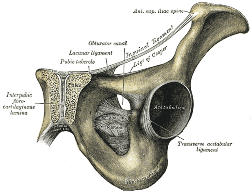
Fibrocartilaginous midline joint between the two pubic bones. It widens in pregnancy and after open-book pelvic injuries, and is a source of groin pain in athletes (osteitis pubis).
Core anatomy
- Location
Anterior midline of the bony pelvis, between the bodies of the two pubic bones.
- Classification
Secondary cartilaginous joint (symphysis)
- Articulating surfaces
The bodies of the right and left pubic bones (parts of the hip bones)
- Capsule
No synovial capsule. The hyaline-covered articular surfaces are joined by a fibrocartilaginous interpubic disc, which may contain a small slit-like cavity.
- Ligaments
- Superior pubic ligament: along the upper border, extending to the pubic tubercles
- Inferior (arcuate) pubic ligament: arches under the joint and rounds off the apex of the pubic arch
- Anterior and posterior fibers blend with the aponeuroses of the abdominal and adductor muscles
- Movements
- Minimal; slight shearing and separation, increased in pregnancy
- Stability
Maintained by the interpubic disc and ligaments. Hormonal ligamentous laxity (relaxin) widens it slightly in late pregnancy.
- Innervation
Branches of the iliohypogastric, ilioinguinal and pudendal nerves verify
- Blood supply
Branches of the obturator, inferior epigastric and internal and external pudendal arteries verify
- Relations
Posterior: the retropubic (prevesical) space and the urinary bladder. Inferior: the dorsal vessels and nerves of the penis or clitoris pass beneath the arcuate ligament. Its upper border is the anterior limit of the pelvic inlet.
Forms a boundary of
Mentioned in
Generated automatically from links in other entries.

Clinical correlations
Symphyseal diastasis (open-book pelvic injury) PANCE
- Mechanism / lesion
Anteroposterior compression of the pelvis, often with sacroiliac ligament disruption.
- Classic presentation
Widened symphysis on AP pelvis, pelvic instability and potential major hemorrhage. Associated with bladder and urethral injuries. See bony pelvis.
Osteitis pubis
- Mechanism / lesion
Repetitive stress at the symphysis from kicking, running and twisting sports, or after pelvic or urologic surgery.
- Classic presentation
Anterior groin and pubic pain, worse with running, kicking or sit-ups, with tenderness over the symphysis. Sclerosis and erosions of the joint margins may appear on X-ray.
Symphysis pubis dysfunction (pregnancy-related pelvic girdle pain)
- Mechanism / lesion
Increased mobility of the pelvic joints in pregnancy.
- Classic presentation
Suprapubic pain worse with walking, climbing stairs, standing on one leg or rolling over in bed. Managed with physical therapy and support belts; true peripartum separation is rare.
High-yield PANCE pearls
- Athlete with midline pubic pain worse with kicking and sit-ups → osteitis pubis.
- Widened symphysis after trauma = open-book (APC) injury: look for hemorrhage and urethral/bladder injury.
Imaging anatomy
- X-ray
AP pelvis: joint width, alignment of the superior pubic rami (step-off in instability) and sclerosis or erosions. Flamingo (single-leg stance) views can show dynamic instability (a vertical shift of more than about 2 mm) verify.
- CT
Defines fractures and diastasis in trauma.
- MRI
Shows bone marrow edema of the pubic bodies (osteitis pubis) and adductor or rectus abdominis aponeurosis injury in athletic groin pain.
- Key views & landmarks
- AP pelvis
- Flamingo views (standing on each leg)
Physical exam
- Surface landmarks
The pubic crest and tubercles are palpable just above the genitalia; the symphysis lies in the midline between them.
- Palpation
Direct tenderness over the joint (osteitis pubis) versus tenderness at the adductor longus origin just lateral to it.
- Strength / function testing
Pain on resisted hip adduction (squeeze test) and on resisted sit-up in athletic groin pain.
Adductor squeeze test
- Technique
Supine with the hips and knees flexed; the patient squeezes the examiner's fist (or a pressure cuff) between the knees.
- Positive test
Reproduction of groin or pubic pain, or reduced squeeze strength.
- Significance
Supports adductor-related or pubic-related groin pain; not specific for osteitis pubis.
Self-quiz
The pubic symphysis is which type of joint?
Correct answer: C
The pubic symphysis is a secondary cartilaginous joint: hyaline cartilage on the bone ends joined by a fibrocartilaginous disc, like the intervertebral discs. A primary cartilaginous joint is a growth plate.
Show answer
C
The pubic symphysis is a secondary cartilaginous joint: hyaline cartilage on the bone ends joined by a fibrocartilaginous disc, like the intervertebral discs. A primary cartilaginous joint is a growth plate.
A 22-year-old soccer player has 6 weeks of midline pubic pain that worsens with kicking and sit-ups. There is tenderness directly over the pubic symphysis. MRI shows bone marrow edema of both pubic bodies. What is the most likely diagnosis?
Correct answer: B
Repetitive shear stress in kicking sports causes osteitis pubis: pain and tenderness at the symphysis with parasymphyseal marrow edema. Femoral neck stress fractures cause groin pain with tenderness over the hip and pain on hip range of motion.
Show answer
B
Repetitive shear stress in kicking sports causes osteitis pubis: pain and tenderness at the symphysis with parasymphyseal marrow edema. Femoral neck stress fractures cause groin pain with tenderness over the hip and pain on hip range of motion.
A pregnant woman at 32 weeks has suprapubic pain that is worse when climbing stairs, standing on one leg and rolling over in bed. What is the most likely cause?
Correct answer: E
Increased mobility of the pelvic joints in pregnancy (relaxin) causes symphysis pubis dysfunction: suprapubic pain with walking, stairs, single-leg stance or rolling over. It is managed with physical therapy and support belts. Round ligament pain is brief, sharp groin pain with sudden movement.
Show answer
E
Increased mobility of the pelvic joints in pregnancy (relaxin) causes symphysis pubis dysfunction: suprapubic pain with walking, stairs, single-leg stance or rolling over. It is managed with physical therapy and support belts. Round ligament pain is brief, sharp groin pain with sudden movement.