Sternum
Flat bone of the anterior thoracic wall made of the manubrium, body and xiphoid process. The sternal angle is the key surface landmark of the thorax.
Core anatomy
- Location
Midline of the anterior thoracic wall.
- Classification
Flat bone.
- Key features & landmarks
- Manubrium: jugular (suprasternal) notch; clavicular notches (sternoclavicular joints); articulates with the 1st costal cartilage
- Sternal angle (of Louis): manubriosternal joint; the 2nd costal cartilage articulates here
- Body: articulates with costal cartilages 3–7 (the 2nd at the sternal angle, the 7th at the xiphisternal junction)
- Xiphoid process: cartilaginous in youth, ossifies with age; xiphisternal joint at about the level of T9
- Relations
Posterior to the manubrium lie the thymus (or its remnant), the left brachiocephalic vein and the arch of the aorta with its branches. Posterior to the body lie the pericardium and the heart, mainly the right ventricle. The internal thoracic artery descends about 1 cm lateral to each sternal border.
- Notes
The sternal angle marks a horizontal plane at the level of the T4/T5 intervertebral disc, which passes through: the 2nd costal cartilage (for counting ribs and intercostal spaces); the boundary between the superior and inferior mediastinum; the beginning and end of the aortic arch; the bifurcation of the trachea (carina, about T4–T5); the bifurcation of the pulmonary trunk; and the point where the azygos vein arches into the superior vena cava. verify
Muscle attachments
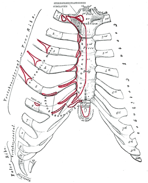
- Pectoralis major — origin
- Sternocleidomastoid — origin
Forms a boundary of
Mentioned in
Generated automatically from links in other entries.
Swipe sideways to see the whole diagram →

Clinical correlations
Sternal fracture PANCE
- Mechanism / lesion
Blunt anterior chest trauma, classically a steering wheel or seatbelt in a deceleration injury.
- Classic presentation
Anterior chest pain and point tenderness, possibly with a palpable step. The main concern is blunt cardiac injury (myocardial contusion, arrhythmia): obtain an ECG (and troponin) and evaluate for other intrathoracic injuries.
Median sternotomy
- Mechanism / lesion
Surgical splitting of the sternum in the midline for cardiac surgery.
- Classic presentation
Complications include sternal wound infection, mediastinitis and sternal dehiscence ("clicking" sternum).
Chest compressions (CPR) PANCE
- Mechanism / lesion
Compression of the heart between the sternum and the vertebral column.
- Classic presentation
Hands are placed on the lower half of the sternum (center of the chest). Pressing on the xiphoid can injure the liver. Sternal and rib fractures are common complications of effective CPR.
High-yield PANCE pearls
- Sternal angle = 2nd rib = T4/T5: aortic arch starts and ends, carina, superior/inferior mediastinum boundary, azygos enters SVC.
- Sternal fracture → screen for blunt cardiac injury with an ECG.
- Count intercostal spaces from the 2nd costal cartilage at the sternal angle.
Imaging anatomy
- X-ray
The sternum is poorly seen on a PA chest film (superimposed on the mediastinum). A lateral sternal view shows fractures and displacement.
- CT
CT (with sagittal reformats) is the most sensitive for sternal fractures and retrosternal hematoma, and evaluates sternal wound infection after sternotomy.
- Ultrasound
Point-of-care ultrasound can detect cortical disruption of the sternum and assess for pericardial effusion.
- Key views & landmarks
- Lateral sternal radiograph
- CT chest with sagittal reformats
Physical exam
- Surface landmarks
Jugular notch (at about the level of the T2/T3 vertebrae), sternal angle (a palpable transverse ridge about 5 cm below the notch), body, and xiphoid process in the epigastric angle. verify
- Palpation
Slide the fingers down from the jugular notch to the ridge of the sternal angle, then move laterally onto the 2nd costal cartilage. The space below it is the 2nd intercostal space. Count down from there.
Self-quiz
Which structure articulates with the sternum at the level of the sternal angle?
Correct answer: B
The 2nd costal cartilage articulates at the manubriosternal joint (sternal angle), so it's the starting point for counting ribs. The 1st costal cartilage joins the manubrium below the clavicle.
Show answer
B
The 2nd costal cartilage articulates at the manubriosternal joint (sternal angle), so it's the starting point for counting ribs. The 1st costal cartilage joins the manubrium below the clavicle.
A horizontal plane through the sternal angle passes through which vertebral level?
Correct answer: B
The sternal (transverse thoracic) plane passes through the T4/T5 disc. It separates the superior and inferior mediastinum and marks the beginning and end of the aortic arch and roughly the tracheal bifurcation.
Show answer
B
The sternal (transverse thoracic) plane passes through the T4/T5 disc. It separates the superior and inferior mediastinum and marks the beginning and end of the aortic arch and roughly the tracheal bifurcation.
A driver in a head-on collision has anterior chest pain and a palpable step-off over the mid-sternum. Which initial test is most important to evaluate a potentially lethal associated injury?
Correct answer: B
Sternal fractures raise concern for blunt cardiac injury. A 12-lead ECG is the initial screening test (with troponin). A normal ECG and troponin make significant blunt cardiac injury unlikely.
Show answer
B
Sternal fractures raise concern for blunt cardiac injury. A 12-lead ECG is the initial screening test (with troponin). A normal ECG and troponin make significant blunt cardiac injury unlikely.
The xiphisternal joint lies at about which vertebral level?
Correct answer: C
The xiphisternal joint lies at about T9. The sternal angle marks the T4/T5 disc; the jugular notch lies at about the level of T2.
Show answer
C
The xiphisternal joint lies at about T9. The sternal angle marks the T4/T5 disc; the jugular notch lies at about the level of T2.
Where should the hands be placed for chest compressions during CPR?
Correct answer: A
Compressions are delivered on the lower half of the sternum, compressing the heart between the sternum and vertebral column. Pressing on the xiphoid can injure the liver. Rib and sternal fractures are common with effective CPR.
Show answer
A
Compressions are delivered on the lower half of the sternum, compressing the heart between the sternum and vertebral column. Pressing on the xiphoid can injure the liver. Rib and sternal fractures are common with effective CPR.