Main bronchi
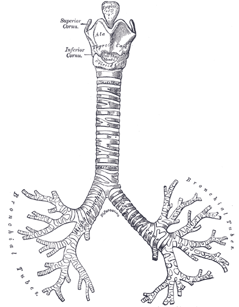
The two airways arising at the carina. The right main bronchus is wider, shorter and more vertical, so foreign bodies and aspirate usually enter the right lung.
Core anatomy
- Location
From the carina (about T4–T5) to the hilum of each lung.
- Parts
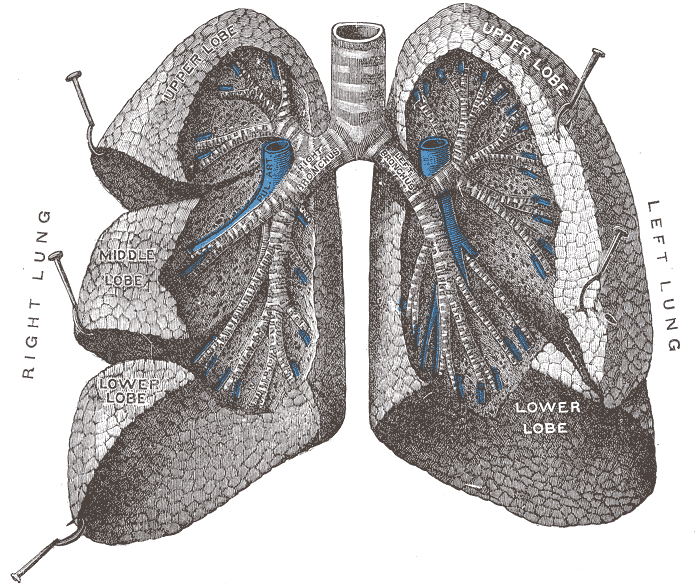
- Right main bronchus: wider, shorter (about 2.5 cm), more vertical; gives the right superior lobar bronchus before entering the hilum, then divides into middle and inferior lobar bronchi
- Left main bronchus: narrower, longer (about 5 cm), more horizontal; passes inferior to the arch of the aorta and anterior to the esophagus and thoracic aorta; divides into superior and inferior lobar bronchi
- Lobar (secondary) bronchi → segmental (tertiary) bronchi, each supplying a bronchopulmonary segment
- Relations
The azygos vein arches over the right main bronchus to reach the superior vena cava. The arch of the aorta passes over the left main bronchus. Subcarinal lymph nodes lie below the carina, and the left atrium lies posteroinferior to the carina.
- Arterial supply
Bronchial arteries (usually one right, two left; from the thoracic aorta or an intercostal artery). verify
- Innervation
Pulmonary plexus (vagus and sympathetic).


Clinical correlations
Foreign body aspiration PANCE
- Mechanism / lesion
Because the right main bronchus is wider and more vertical, inhaled objects most often lodge on the right. Common in toddlers (nuts, small toys).
- Classic presentation
Sudden choking and coughing, then unilateral wheeze, decreased breath sounds or recurrent pneumonia on one side. Unilateral hyperinflation (air trapping) on expiratory or decubitus films if the object acts as a check valve.
Bronchogenic carcinoma (central) PANCE
- Mechanism / lesion
Squamous cell and small cell carcinomas typically arise in central (main/lobar) bronchi.
- Classic presentation
Cough, hemoptysis, postobstructive pneumonia or lobar collapse, and hilar mass on imaging.
Tracheobronchial injury
- Mechanism / lesion
Blunt chest trauma with rapid deceleration, usually within about 2.5 cm of the carina.
- Classic presentation
Pneumomediastinum, subcutaneous emphysema, and a persistent large air leak despite a chest tube. The collapsed lung falls away from the hilum ("fallen lung" sign).
High-yield PANCE pearls
- Right main bronchus: wider, shorter, more vertical → foreign bodies and aspiration go right.
- Child with sudden cough and unilateral wheeze → foreign body; bronchoscopy for diagnosis and removal.
- Air trapping on expiratory films → check-valve foreign body.
Imaging anatomy
- X-ray
Most aspirated foreign bodies are radiolucent. Look for indirect signs: unilateral hyperinflation (air trapping, best seen on expiratory or lateral decubitus films), atelectasis, or postobstructive pneumonia.
- CT
Defines endobronchial lesions, central tumors, bronchiectasis and airway injury. Virtual bronchoscopy is possible.
- Key views & landmarks
- Inspiratory and expiratory chest films (or bilateral decubitus in young children)
- Bronchoscopy (rigid for foreign body removal) — diagnostic and therapeutic
Physical exam
- Surface landmarks
The bronchi lie deep behind the sternal angle and upper sternum. Bronchovesicular breath sounds are normally heard over the main bronchi (1st–2nd intercostal spaces anteriorly and between the scapulae posteriorly).
- Strength / function testing
Compare breath sounds and wheeze side to side. A unilateral, fixed wheeze suggests a focal airway obstruction.
Self-quiz
A 2-year-old choked while eating peanuts. He now has a persistent cough and wheezing over one side of the chest. Where is the foreign body most likely lodged?
Correct answer: B
The right main bronchus is wider, shorter and more vertical, so aspirated foreign bodies lodge there most often. Confirm and remove with rigid bronchoscopy.
Show answer
B
The right main bronchus is wider, shorter and more vertical, so aspirated foreign bodies lodge there most often. Confirm and remove with rigid bronchoscopy.
Which structure arches over the right main bronchus?
Correct answer: B
The azygos vein arches over the right main bronchus (root of the right lung) at about T4 to enter the SVC. The aortic arch passes over the left main bronchus.
Show answer
B
The azygos vein arches over the right main bronchus (root of the right lung) at about T4 to enter the SVC. The aortic arch passes over the left main bronchus.
A child who aspirated a small toy has a chest radiograph on expiration showing the left lung hyperlucent while the right lung decreases in volume normally. What does this indicate?
Correct answer: B
Air enters the left lung around the object during inspiration but can't leave on expiration (check-valve), so that side stays hyperinflated while the normal lung deflates. Foreign bodies occur on the left too, even though the right is more common.
Show answer
B
Air enters the left lung around the object during inspiration but can't leave on expiration (check-valve), so that side stays hyperinflated while the normal lung deflates. Foreign bodies occur on the left too, even though the right is more common.
The left main bronchus passes inferior to which structure?
Correct answer: B
The arch of the aorta passes over the left main bronchus, while the azygos vein arches over the right main bronchus. The left main bronchus is longer and more horizontal than the right.
Show answer
B
The arch of the aorta passes over the left main bronchus, while the azygos vein arches over the right main bronchus. The left main bronchus is longer and more horizontal than the right.
After a high-speed deceleration injury, a patient has pneumomediastinum and a persistent large air leak despite a functioning chest tube. The collapsed lung has fallen away from the hilum. What is the most likely injury?
Correct answer: C
Tracheobronchial injury, usually within about 2.5 cm of the carina, causes pneumomediastinum, subcutaneous emphysema, a persistent air leak despite a chest tube, and the "fallen lung" sign.
Show answer
C
Tracheobronchial injury, usually within about 2.5 cm of the carina, causes pneumomediastinum, subcutaneous emphysema, a persistent air leak despite a chest tube, and the "fallen lung" sign.