Right coronary artery
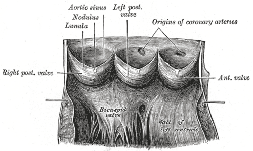
Arises from the right aortic sinus and runs in the right atrioventricular groove. It usually supplies the SA and AV nodes, the right ventricle and (in right dominance) the inferior wall; occlusion causes inferior MI.
Core anatomy
- Location
Right atrioventricular (coronary) groove, then the posterior interventricular groove (in right-dominant hearts).
- Origin
Right aortic sinus of the ascending aorta (above the right cusp of the aortic valve).
- Course
Passes between the right auricle and the pulmonary trunk, descends in the right atrioventricular groove around the acute margin of the heart to the posterior surface. In a right-dominant circulation (most people) it gives the posterior interventricular (posterior descending) artery at the crux.
- Branches
- SA nodal artery (in about 60%)
- Conus artery (to the RV outflow tract)
- Right marginal (acute marginal) artery: along the inferior (acute) margin, supplying the RV
- AV nodal artery (from the dominant artery, usually the RCA)
- Posterior interventricular (posterior descending) artery: in right dominance; supplies the posterior third of the septum and the inferior LV wall, and the posteromedial papillary muscle
- Posterolateral branches to the LV (in right dominance)
- Supplies
Right atrium, most of the right ventricle, SA node (about 60%), AV node (about 80%), the posterior third of the interventricular septum and the inferior (diaphragmatic) LV wall (in right dominance), and the posteromedial papillary muscle. verify
- Anastomoses
Small anastomoses with branches of the left coronary artery (e.g. septal and apical collaterals), usually inadequate for sudden occlusion.
- Notes
Coronary dominance is defined by which artery gives the posterior interventricular artery: right dominant in most people (roughly 70–80%), left dominant (from the circumflex artery) in a minority, and codominant in the rest. verify
Structures supplied
Mentioned in
Generated automatically from links in other entries.

Clinical correlations
Inferior myocardial infarction PANCE
- Mechanism / lesion
Occlusion of the RCA (in right-dominant circulation).
- Classic presentation
ST elevation in II, III and aVF, with reciprocal ST depression in I and aVL. Bradycardia and AV block (nodal ischemia). Possible posteromedial papillary muscle rupture (acute MR).
Right ventricular infarction PANCE
- Mechanism / lesion
Proximal RCA occlusion (before the acute marginal branches) with an inferior MI.
- Classic presentation
Hypotension, clear lungs, elevated JVP (the preload-dependent RV fails). ST elevation in right-sided leads (V4R). Avoid nitrates and other preload reducers; give IV fluids.
High-yield PANCE pearls
- Inferior MI: II, III, aVF → RCA (usually).
- Inferior MI + hypotension + clear lungs + JVD → RV infarct: get V4R; avoid nitrates, give fluids.
- RCA usually supplies the SA and AV nodes → bradycardia and heart block.
- Right dominance = posterior interventricular artery comes from the RCA.
Imaging anatomy
- CT
Coronary CT angiography shows stenosis, plaque, anomalous origin (e.g. an RCA from the left sinus coursing between the aorta and pulmonary trunk, a cause of sudden death in young athletes), and dominance.
- MRI
Late gadolinium enhancement localizes infarcted territory.
- Ultrasound
Echo shows regional wall motion abnormalities of the inferior wall and RV free wall in RCA ischemia.
- Key views & landmarks
- Invasive coronary angiography: the definitive study (LAO view shows the RCA well)
- 12-lead ECG with right-sided leads (V4R)
Physical exam
- Strength / function testing
In inferior MI, check heart rate and rhythm (bradycardia, AV block), JVP and lung fields (RV infarct), and listen for a new holosystolic murmur (papillary muscle rupture).
Self-quiz
A 64-year-old man with inferior ST elevation (II, III, aVF) becomes hypotensive after sublingual nitroglycerin. His lungs are clear and his JVP is elevated. What is the most likely explanation?
Correct answer: B
A proximal RCA occlusion can infarct the right ventricle, which depends on preload. Nitrates reduce preload and cause hypotension. Treat with IV fluids and confirm with right-sided leads (V4R).
Show answer
B
A proximal RCA occlusion can infarct the right ventricle, which depends on preload. Nitrates reduce preload and cause hypotension. Treat with IV fluids and confirm with right-sided leads (V4R).
Coronary dominance is determined by which artery gives rise to which branch?
Correct answer: C
Dominance is defined by the origin of the posterior interventricular artery: the RCA in right dominance (most people) and the circumflex in left dominance.
Show answer
C
Dominance is defined by the origin of the posterior interventricular artery: the RCA in right dominance (most people) and the circumflex in left dominance.
Which ECG leads show ST elevation in an inferior wall MI?
Correct answer: D
II, III and aVF face the inferior wall, usually supplied by the RCA. V1–V4 are septal/anterior (LAD); I, aVL, V5–V6 are lateral (circumflex).
Show answer
D
II, III and aVF face the inferior wall, usually supplied by the RCA. V1–V4 are septal/anterior (LAD); I, aVL, V5–V6 are lateral (circumflex).
Which branch of the RCA runs along the inferior (acute) margin of the heart to supply the right ventricle?
Correct answer: C
The right (acute) marginal artery runs along the acute margin and supplies the RV. Obtuse marginals come from the circumflex, and diagonals from the LAD.
Show answer
C
The right (acute) marginal artery runs along the acute margin and supplies the RV. Obtuse marginals come from the circumflex, and diagonals from the LAD.
In an inferior STEMI, in which leads is reciprocal ST depression usually seen?
Correct answer: D
Inferior MI (II, III, aVF) typically shows reciprocal ST depression in I and aVL. ST elevation in V4R suggests right ventricular involvement.
Show answer
D
Inferior MI (II, III, aVF) typically shows reciprocal ST depression in I and aVL. ST elevation in V4R suggests right ventricular involvement.