Ventricular system and CSF
Four interconnected cavities in the brain that produce and circulate cerebrospinal fluid. Blockage or impaired absorption causes hydrocephalus, including normal pressure hydrocephalus in older adults.
Core anatomy
- Location
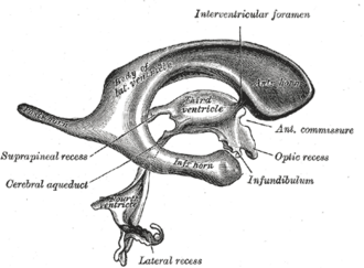
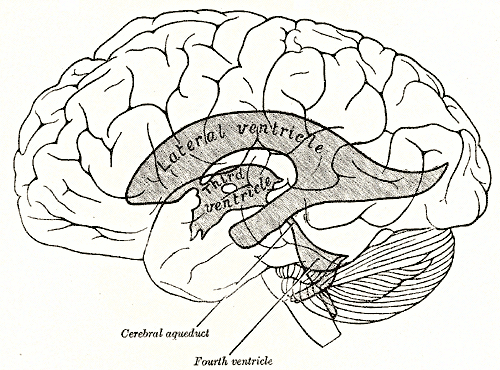
Within the brain — two lateral ventricles in the cerebral hemispheres, the third ventricle between the thalami, the cerebral aqueduct through the midbrain and the fourth ventricle between the pons/medulla and cerebellum.
- Boundaries
- lateral ventricles
- Frontal horn, body, occipital horn and temporal horn (the hippocampus lies in its floor); the caudate forms the lateral wall of the body
- third ventricle
- Thalami (lateral walls), hypothalamus (floor), lamina terminalis (front)
- fourth ventricle
- Pons and medulla (floor, rhomboid fossa), cerebellum (roof)
- Contents
- CSF — about 150 mL total, produced at about 500 mL/day (replaced 3–4 times daily), mainly by the choroid plexus (lateral, third and fourth ventricles)
- Choroid plexus (ependymal cells over capillaries)
- Communications
Lateral ventricles → interventricular foramina (of Monro) → third ventricle → cerebral aqueduct (of Sylvius) → fourth ventricle → median aperture (Magendie) and two lateral apertures (Luschka) → subarachnoid space and cisterns (and the central canal of the cord) → absorbed through arachnoid granulations into the superior sagittal sinus (see dural venous sinuses, cranial meninges and intracranial hemorrhage).
- Notes
CSF cushions the brain (buoyancy), removes metabolites and maintains a stable chemical environment. Normal opening pressure on lumbar puncture is about 6–20 cm H₂O (lateral decubitus). verify
Mentioned in
Generated automatically from links in other entries.
Swipe sideways to see the whole diagram →


Clinical correlations
Hydrocephalus PANCE
- Mechanism / lesion
Noncommunicating (obstructive) — blockage within the ventricles: aqueductal stenosis, tumors (posterior fossa, colloid cyst of the third ventricle), Chiari II/Dandy–Walker. Communicating — impaired absorption at the arachnoid granulations: after subarachnoid hemorrhage or meningitis; normal pressure hydrocephalus.
- Classic presentation
Adults: headache (worse in the morning), vomiting, papilledema, declining consciousness, CN VI palsy. Infants: enlarging head circumference, bulging fontanelle, splayed sutures, "sunset eyes" (upgaze palsy). Treat with a ventriculoperitoneal shunt or endoscopic third ventriculostomy; acute cases need an external ventricular drain.
Normal pressure hydrocephalus PANCE
- Mechanism / lesion
Communicating hydrocephalus in older adults, often idiopathic; normal opening pressure on LP.
- Classic presentation
Triad "wet, wobbly and wacky": urinary incontinence, gait apraxia (magnetic, shuffling, wide-based gait — usually first), and dementia. CT/MRI: ventricles enlarged out of proportion to atrophy. Improvement after large-volume lumbar puncture predicts benefit from a VP shunt — a reversible dementia.
Colloid cyst of the third ventricle
- Mechanism / lesion
Benign cyst near the foramen of Monro that can intermittently block CSF outflow.
- Classic presentation
Positional headaches, drop attacks and sudden death from acute hydrocephalus.
High-yield PANCE pearls
- Gait apraxia + incontinence + dementia → NPH → large-volume LP → VP shunt.
- Infant with rapidly enlarging head, bulging fontanelle and sunset eyes → hydrocephalus → ultrasound through the fontanelle.
- Hydrocephalus after SAH or meningitis → communicating (arachnoid granulations).
- CSF path: lateral → Monro → third → aqueduct → fourth → Luschka/Magendie → subarachnoid → arachnoid granulations → superior sagittal sinus.
Imaging anatomy
- CT
Noncontrast CT — ventricular size, periventricular edema (transependymal flow), shunt problems.
- MRI
MRI — cause of obstruction (aqueductal stenosis, tumor), CSF flow studies, NPH (enlarged ventricles, narrow high-convexity sulci).
- Ultrasound
Cranial ultrasound through the anterior fontanelle in infants.
- Key views & landmarks
- Shunt series (X-rays along the tubing) for suspected shunt malfunction
Physical exam
- Surface landmarks
Lumbar puncture below the conus — at L3–L4 or L4–L5 (the iliac crests mark about L4; see spinal meninges and spaces).
- Strength / function testing
Head circumference (plot on growth charts), fontanelle, fundi (papilledema), gait (NPH: timed walk before and after LP), cognition; examine for shunt malfunction (headache, vomiting, lethargy).
Self-quiz
A 74-year-old man has had progressive urinary urgency and incontinence, forgetfulness, and a slow, shuffling gait in which his feet seem stuck to the floor. CT shows enlarged ventricles without proportional sulcal widening. What is the most appropriate next step?
Correct answer: B
This is normal pressure hydrocephalus (wet, wobbly, wacky). Clinical improvement after large-volume CSF removal supports a VP shunt, which can reverse symptoms.
Show answer
B
This is normal pressure hydrocephalus (wet, wobbly, wacky). Clinical improvement after large-volume CSF removal supports a VP shunt, which can reverse symptoms.
An MRI shows dilated lateral and third ventricles and a normal-sized fourth ventricle. Where is the obstruction most likely?
Correct answer: B
Dilation proximal to the aqueduct (lateral and third ventricles) with a normal fourth ventricle localizes the block to the cerebral aqueduct (aqueductal stenosis).
Show answer
B
Dilation proximal to the aqueduct (lateral and third ventricles) with a normal fourth ventricle localizes the block to the cerebral aqueduct (aqueductal stenosis).
Where is most cerebrospinal fluid produced?
Correct answer: C
CSF (about 500 mL/day, about 150 mL at a time) is made mainly by the choroid plexus of the lateral, third and fourth ventricles and absorbed through arachnoid granulations into the superior sagittal sinus.
Show answer
C
CSF (about 500 mL/day, about 150 mL at a time) is made mainly by the choroid plexus of the lateral, third and fourth ventricles and absorbed through arachnoid granulations into the superior sagittal sinus.
Hydrocephalus after subarachnoid hemorrhage or meningitis is usually caused by what?
Correct answer: D
Blood or pus impairs absorption at the arachnoid granulations, causing communicating hydrocephalus. Blocks within the ventricular system cause noncommunicating hydrocephalus.
Show answer
D
Blood or pus impairs absorption at the arachnoid granulations, causing communicating hydrocephalus. Blocks within the ventricular system cause noncommunicating hydrocephalus.
Which finding in an infant suggests hydrocephalus?
Correct answer: B
Infant hydrocephalus causes rapidly enlarging head circumference, a bulging fontanelle, splayed sutures and "sunset eyes" (upgaze palsy). Ultrasound through the fontanelle is the first test.
Show answer
B
Infant hydrocephalus causes rapidly enlarging head circumference, a bulging fontanelle, splayed sutures and "sunset eyes" (upgaze palsy). Ultrasound through the fontanelle is the first test.