Liver
Largest gland in the body, in the right upper quadrant. It has a dual blood supply (portal vein and hepatic artery) and is divided into eight functional segments; cirrhosis and portal hypertension are its central clinical problems.
Core anatomy
- Location
Right upper quadrant and epigastrium, under the right dome of the diaphragm, protected by the lower ribs.
- Parts
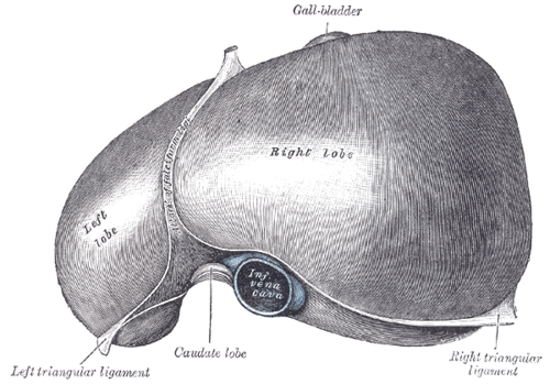
- Anatomical lobes: right, left, caudate (posterior, between the IVC and ligamentum venosum) and quadrate (inferior, between the gallbladder and ligamentum teres)
- Functional (surgical) lobes: right and left, separated by Cantlie line (from the gallbladder fossa to the IVC). Divided into eight Couinaud segments, each with its own portal pedicle and biliary drainage, allowing segmental resection
- Porta hepatis: entry of the hepatic portal vein and hepatic artery proper; exit of the hepatic ducts
- Bare area: on the posterior surface, in direct contact with the diaphragm (no peritoneum)
- Peritoneal ligaments: falciform ligament (with the ligamentum teres, the remnant of the umbilical vein, in its free edge), coronary and triangular ligaments, lesser omentum (hepatogastric and hepatoduodenal ligaments)
- Relations
Diaphragmatic surface under the diaphragm. Visceral surface contacts the stomach, duodenum, hepatic flexure of the colon, right kidney and suprarenal gland, and the gallbladder and biliary tree. The inferior vena cava runs in a groove on the posterior surface, receiving the hepatic veins.
- Arterial supply
Dual supply: the portal vein (about 75% of inflow; nutrient-rich, relatively deoxygenated) and the hepatic artery proper (about 25%; oxygenated) from the common hepatic artery (celiac trunk). verify
- Venous drainage
Right, middle and left hepatic veins → inferior vena cava just below the diaphragm.
- Lymphatic drainage
Hepatic nodes at the porta hepatis → celiac nodes; some drain through the diaphragm to thoracic nodes.
- Innervation
Hepatic plexus (vagus and sympathetic). The capsule (Glisson) is sensitive to stretch, so hepatomegaly causes RUQ discomfort; diaphragmatic irritation can refer to the right shoulder.
Forms a boundary of
Mentioned in
Generated automatically from links in other entries.
Swipe sideways to see the whole diagram →

Clinical correlations
Cirrhosis and portal hypertension PANCE
- Mechanism / lesion
Chronic injury (alcohol, viral hepatitis B/C, MASLD/NAFLD, hemochromatosis, autoimmune disease) leads to fibrosis and nodular regeneration, raising resistance to portal flow.
- Classic presentation
Portal hypertension: ascites, splenomegaly, esophageal varices, caput medusae. Liver failure: jaundice, coagulopathy (raised INR), hypoalbuminemia, hepatic encephalopathy (asterixis), spider angiomas, palmar erythema, gynecomastia.
Hepatocellular carcinoma PANCE
- Mechanism / lesion
Usually in a cirrhotic liver or with chronic hepatitis B (even without cirrhosis).
- Classic presentation
Often found on surveillance ultrasound (every 6 months in at-risk patients, ± AFP). Decompensation of known cirrhosis, weight loss, RUQ mass. Diagnosed on multiphase CT or MRI (arterial enhancement with washout).
Liver injury in trauma PANCE
- Mechanism / lesion
Large, relatively fixed solid organ; injured in both blunt and penetrating trauma.
- Classic presentation
RUQ pain, hemoperitoneum (positive FAST), shock. Stable patients are often managed non-operatively after CT grading.
Budd–Chiari syndrome
- Mechanism / lesion
Hepatic venous outflow obstruction (thrombosis), associated with hypercoagulable states (e.g. polycythemia vera, pregnancy, OCPs).
- Classic presentation
Triad of abdominal pain, ascites and hepatomegaly. The caudate lobe may enlarge (it drains directly to the IVC).
High-yield PANCE pearls
- Dual supply: portal vein ~75%, hepatic artery ~25%.
- Cirrhosis complications: ascites, varices, encephalopathy, SBP, HCC, hepatorenal syndrome.
- HCC surveillance in cirrhosis: ultrasound every 6 months ± AFP.
- Budd–Chiari: pain + ascites + hepatomegaly; hypercoagulable state.
- Ligamentum teres = remnant of the umbilical vein; ligamentum venosum = remnant of the ductus venosus.
Imaging anatomy
- X-ray
Not useful for the liver itself. Calcifications or gas in the portal venous system (a sign of bowel ischemia) may be seen.
- CT
Multiphase (arterial, portal venous, delayed) CT characterizes liver lesions (HCC: arterial hyperenhancement and washout), shows trauma injuries, and features of cirrhosis (nodular contour, caudate hypertrophy, splenomegaly, varices, ascites).
- MRI
MRI (with liver-specific contrast) is the most accurate for characterizing focal lesions. Quantifies iron and fat.
- Ultrasound
First-line for RUQ pain, abnormal liver tests, and HCC surveillance. Cirrhotic liver: coarse echotexture and nodular surface. Doppler assesses portal vein flow direction and patency and hepatic vein patency (Budd–Chiari). Elastography estimates fibrosis.
- Key views & landmarks
- RUQ ultrasound with Doppler
- Multiphase CT or MRI for focal lesions
Physical exam
- Surface landmarks
Upper border at about the 5th intercostal space in the right midclavicular line; lower edge at or just below the right costal margin. Normal liver span by percussion in the right midclavicular line is roughly 6–12 cm. verify
- Palpation
Place the hand below the right costal margin (or start from the RLQ if hepatomegaly is suspected) and ask the patient to breathe in deeply; feel for the edge descending onto the fingers. Note consistency (firm, nodular), tenderness and pulsatility.
- Strength / function testing
Percussion from resonant lung downward to dullness (upper border) and from tympanic abdomen upward (lower border) in the midclavicular line. The scratch test is an alternative for locating the lower edge.
Self-quiz
What proportion of hepatic blood flow is supplied by the portal vein?
Correct answer: D
The portal vein provides about 75% of hepatic blood flow; the hepatic artery provides about 25% but a large share of the oxygen.
Show answer
D
The portal vein provides about 75% of hepatic blood flow; the hepatic artery provides about 25% but a large share of the oxygen.
A 34-year-old woman with polycythemia vera develops RUQ pain, hepatomegaly and ascites over two weeks. Doppler ultrasound shows no flow in the hepatic veins. What is the diagnosis?
Correct answer: B
Budd–Chiari syndrome (hepatic vein thrombosis) causes the triad of pain, ascites and hepatomegaly and is associated with myeloproliferative disorders such as polycythemia vera.
Show answer
B
Budd–Chiari syndrome (hepatic vein thrombosis) causes the triad of pain, ascites and hepatomegaly and is associated with myeloproliferative disorders such as polycythemia vera.
A patient with cirrhosis should undergo which surveillance for hepatocellular carcinoma?
Correct answer: B
Patients with cirrhosis (and some with chronic hepatitis B) should have ultrasound every 6 months, with or without AFP.
Show answer
B
Patients with cirrhosis (and some with chronic hepatitis B) should have ultrasound every 6 months, with or without AFP.
The functional right and left lobes of the liver are separated by a plane from the gallbladder fossa to which structure?
Correct answer: B
Cantlie line runs from the gallbladder fossa to the IVC, following the middle hepatic vein. The falciform ligament marks the anatomical (not functional) division.
Show answer
B
Cantlie line runs from the gallbladder fossa to the IVC, following the middle hepatic vein. The falciform ligament marks the anatomical (not functional) division.
The ligamentum teres in the free edge of the falciform ligament is the remnant of which fetal structure?
Correct answer: B
The ligamentum teres is the remnant of the umbilical vein; the ligamentum venosum is the remnant of the ductus venosus. The paraumbilical veins run with the ligamentum teres, which is how caput medusae forms.
Show answer
B
The ligamentum teres is the remnant of the umbilical vein; the ligamentum venosum is the remnant of the ductus venosus. The paraumbilical veins run with the ligamentum teres, which is how caput medusae forms.