Hip joint
Deep ball-and-socket joint between the femoral head and the acetabulum, built for stability. Osteoarthritis, posterior dislocation, septic arthritis in children and developmental dysplasia are the high-yield topics.
Core anatomy
- Location
Between the head of the femur and the acetabulum of the hip bone, deep to the femoral triangle anteriorly and the gluteal muscles posteriorly.
- Classification
Synovial ball-and-socket (spheroidal) joint
- Capsule
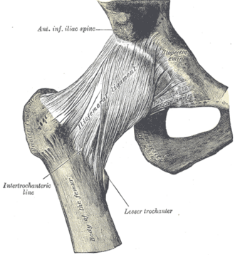
Strong; attached to the acetabular rim and, on the femur, to the intertrochanteric line in front but only halfway down the neck behind. The acetabular labrum (fibrocartilage) deepens the socket.
- Ligaments
- Iliofemoral ligament (Y-shaped, of Bigelow): the strongest ligament in the body; prevents hyperextension, letting us stand with little muscle effort
- Pubofemoral ligament: limits abduction
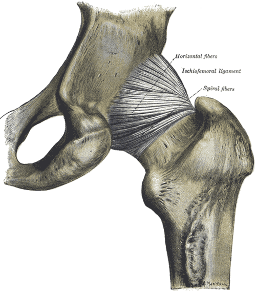
- Ischiofemoral ligament: posterior; the weakest
- Ligament of the head of the femur: carries a small artery (from the obturator artery), important in children
- Transverse acetabular ligament: bridges the acetabular notch
- Movements
- Flexion (iliopsoas, rectus femoris, sartorius)
- Extension (gluteus maximus, hamstrings)
- Abduction (gluteus medius and minimus)
- Adduction (adductor muscles of the thigh)
- Medial rotation (anterior fibers of gluteus medius/minimus, tensor fasciae latae)
- Lateral rotation (piriformis, obturators, gemelli, quadratus femoris, gluteus maximus)
- Stability
The deep acetabulum and labrum, the strong capsular ligaments (tight in extension), and surrounding muscles. Most stable in extension; most vulnerable to posterior dislocation when flexed and adducted.
- Innervation
Hilton law — nerves to the muscles crossing it: Femoral nerve, obturator nerve, the nerve to quadratus femoris and the superior gluteal nerve. Hence hip pain is often felt in the groin, thigh or knee.
- Blood supply
Medial and lateral circumflex femoral arteries (medial is the main supply to the femoral head) and the artery of the ligament of the head (obturator).
- Relations
Anteriorly iliopsoas and the femoral nerve and vessels; posteriorly the short lateral rotators and the sciatic nerve.


Clinical correlations
Hip osteoarthritis PANCE
- Mechanism / lesion
Degeneration of articular cartilage with age, prior injury, dysplasia or femoroacetabular impingement.
- Classic presentation
Groin (or anterior thigh/knee) pain worse with activity, morning stiffness under 30 minutes, and loss of internal rotation first. X-ray: joint space narrowing, osteophytes, subchondral sclerosis and cysts. Treat with exercise, weight loss, NSAIDs, then total hip arthroplasty.
Posterior hip dislocation PANCE
- Mechanism / lesion
A dashboard injury — force along the femur with the hip flexed and adducted (the most common hip dislocation, about 90%).
- Classic presentation
The leg is shortened, flexed, adducted and internally rotated. Risks: sciatic nerve injury (fibular division) and AVN of the femoral head — reduce urgently (within about 6 hours). Anterior dislocations present abducted and externally rotated.
Septic arthritis vs transient synovitis in children PANCE
- Mechanism / lesion
Septic arthritis is usually Staphylococcus aureus (Kingella in young children); transient synovitis often follows a viral illness.
- Classic presentation
Limp or refusal to bear weight. Kocher criteria favor septic arthritis: fever over 38.5 °C, non-weight-bearing, ESR over 40 mm/h and WBC over 12,000/µL (plus CRP over 2 mg/dL). Ultrasound shows an effusion; urgent aspiration and washout for septic arthritis.
Developmental dysplasia of the hip (DDH) PANCE
- Mechanism / lesion
Shallow acetabulum and lax capsule; risk factors — breech presentation, female sex, first-born, family history, oligohydramnios.
- Classic presentation
Newborn screening with Barlow and Ortolani tests; later, asymmetric thigh folds, limited abduction, leg-length discrepancy (Galeazzi sign) or a waddling gait. Ultrasound before about 4–6 months, X-ray after; treat with a Pavlik harness.
High-yield PANCE pearls
- Dashboard injury, leg shortened, adducted, internally rotated → posterior hip dislocation (sciatic nerve, AVN).
- Hip OA: groin pain and loss of internal rotation.
- Febrile child refusing to bear weight → consider septic hip (Kocher criteria) → aspirate.
- DDH: breech, female, first-born; Barlow/Ortolani; Pavlik harness.
- Hip pathology often refers pain to the knee.
Imaging anatomy
- X-ray
AP pelvis and lateral hip — OA, fractures, dislocation, dysplasia, SCFE, Perthes.
- MRI
Labral tears (MR arthrography), AVN, occult fractures, osteomyelitis.
- Ultrasound
Hip ultrasound detects effusions (septic arthritis, transient synovitis) and guides aspiration; it is the test for DDH in infants under about 4–6 months (before the femoral head ossifies).
- Key views & landmarks
- AP pelvis + lateral hip
- Infant hip ultrasound (Graf method) for DDH
Physical exam
- Surface landmarks
The joint lies deep to a point about 2–3 cm below the midpoint of the inguinal ligament, lateral to the femoral pulse; the greater trochanter is palpable laterally.
- Palpation
Groin tenderness suggests an intra-articular process; greater trochanteric tenderness suggests gluteal tendinopathy/bursitis.
- Strength / function testing
Range of motion, especially internal rotation with the hip flexed to 90° (reduced early in OA); gait; Trendelenburg test (see gluteus medius and minimus).
FADIR (anterior impingement) test
- Technique
Supine; flex the hip to 90°, then adduct and internally rotate.
- Positive test
Groin pain.
- Significance
Suggests femoroacetabular impingement or a labral tear.
Barlow and Ortolani tests (infants)
- Technique
Barlow: with the hip flexed and adducted, push posteriorly to try to dislocate it. Ortolani: abduct the flexed hip while lifting the greater trochanter forward to relocate it.
- Positive test
A palpable "clunk" as the femoral head dislocates (Barlow) or relocates (Ortolani).
- Significance
Developmental dysplasia of the hip.
Thomas test
- Technique
Supine; the patient holds one knee to the chest (flattening the lumbar lordosis) while the other leg rests on the table.
- Positive test
The thigh of the extended leg lifts off the table.
- Significance
Hip flexion contracture (tight iliopsoas).
Self-quiz
A driver's knee strikes the dashboard in a head-on collision. His right leg is shortened, flexed, adducted and internally rotated. Which nerve is most at risk?
Correct answer: C
This is a posterior hip dislocation. The femoral head is driven backward toward the sciatic nerve (especially its fibular division, causing foot drop). Urgent reduction also reduces the risk of AVN.
Show answer
C
This is a posterior hip dislocation. The femoral head is driven backward toward the sciatic nerve (especially its fibular division, causing foot drop). Urgent reduction also reduces the risk of AVN.
A 3-year-old has a temperature of 39.2 °C and refuses to bear weight on the right leg. ESR is 55 mm/h and WBC is 15,000/µL. Ultrasound shows a hip effusion. What is the most appropriate next step?
Correct answer: B
The child meets all four Kocher criteria (fever, non-weight-bearing, high ESR, high WBC), so septic arthritis is highly likely. Urgent aspiration (and surgical drainage) plus IV antibiotics are needed to prevent joint destruction.
Show answer
B
The child meets all four Kocher criteria (fever, non-weight-bearing, high ESR, high WBC), so septic arthritis is highly likely. Urgent aspiration (and surgical drainage) plus IV antibiotics are needed to prevent joint destruction.
Which ligament of the hip is the strongest and prevents hyperextension?
Correct answer: C
The Y-shaped iliofemoral ligament is the strongest ligament in the body and resists hyperextension, which lets us stand upright with minimal muscle work.
Show answer
C
The Y-shaped iliofemoral ligament is the strongest ligament in the body and resists hyperextension, which lets us stand upright with minimal muscle work.
Which newborn screening tests are used for developmental dysplasia of the hip?
Correct answer: B
Barlow (attempts to dislocate a reduced hip) and Ortolani (reduces a dislocated hip with a clunk) are the newborn screening tests. Risk factors are breech presentation, female sex, being first-born and family history. Ultrasound is used before about 4–6 months and a Pavlik harness is the usual treatment.
Show answer
B
Barlow (attempts to dislocate a reduced hip) and Ortolani (reduces a dislocated hip with a clunk) are the newborn screening tests. Risk factors are breech presentation, female sex, being first-born and family history. Ultrasound is used before about 4–6 months and a Pavlik harness is the usual treatment.
Which hip movement is usually lost first in hip osteoarthritis?
Correct answer: C
Loss of internal rotation is the earliest and most characteristic finding in hip OA, along with groin (or anterior thigh/knee) pain worse with activity.
Show answer
C
Loss of internal rotation is the earliest and most characteristic finding in hip OA, along with groin (or anterior thigh/knee) pain worse with activity.