Femoral nerve
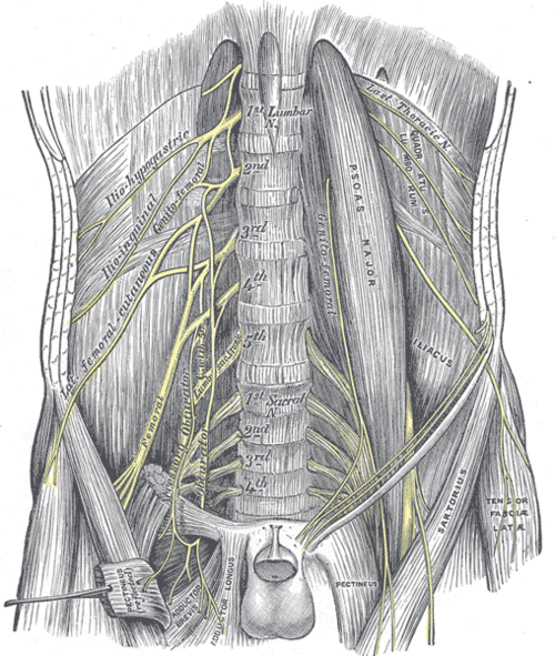
Largest branch of the lumbar plexus (L2–L4), supplying the hip flexors and knee extensors and the skin of the anterior thigh and medial leg. Lesions cause knee buckling and a lost patellar reflex.
Core anatomy
- Location
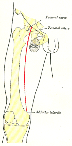
Anterior thigh; enters the thigh under the inguinal ligament lateral to the femoral artery.
- Root values
Posterior divisions of the ventral rami of L2–L4.
- Origin
Lumbar plexus, within psoas major
- Course
Emerges from the lateral border of psoas, runs between psoas and iliacus (supplying iliacus), passes under the inguinal ligament lateral to the femoral sheath (it is not in the sheath) into the femoral triangle, and divides after a few centimeters into anterior and posterior branches.
- Branches
- Muscular: iliacus, pectineus, sartorius and quadriceps femoris
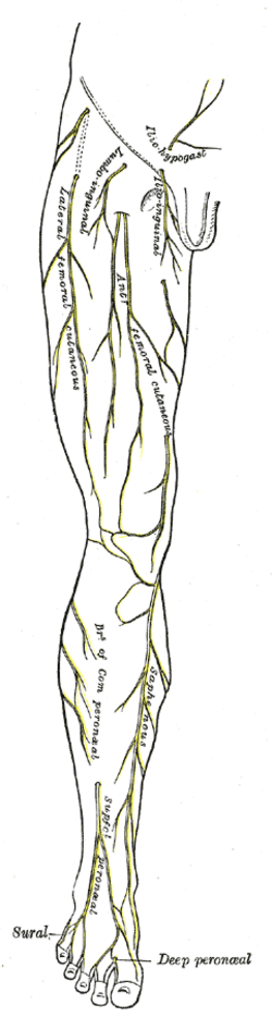
- Anterior cutaneous nerves of the thigh
- Saphenous nerve: the longest cutaneous branch; runs through the adductor canal, emerges at the medial knee, and runs with the great saphenous vein to the medial leg and medial foot
- Articular branches to the hip and knee
- Motor
Hip flexion (iliacus, sartorius, rectus femoris) and knee extension (quadriceps).
- Sensory
Anterior and medial thigh; medial leg and medial foot (saphenous).
- Relations
Order in the femoral triangle from lateral to medial: Nerve, Artery, Vein, (Empty space), Lymphatics — NAVEL.
Muscles innervated
- Adductor muscles of the thigh — obturator nerve (L2–L4) for most; pectineus mainly by the femoral nerve; the hamstring part of adductor magnus by the tibial division of the sciatic nerve
- Quadriceps femoris — femoral nerve (L2–L4)
Other structures innervated
Found in
Mentioned in
Generated automatically from links in other entries.
Swipe sideways to see the whole diagram →
Swipe sideways to see the whole diagram →



Clinical correlations
Femoral neuropathy PANCE
- Mechanism / lesion
Retroperitoneal/iliopsoas hematoma (anticoagulation, hemophilia), pelvic or hip surgery (retractor compression, lithotomy positioning), diabetic amyotrophy, femoral artery catheterization.
- Classic presentation
Weak knee extension (knee buckles, trouble with stairs), weak hip flexion (if the lesion is proximal), absent/reduced patellar reflex, and numbness of the anterior thigh and medial leg. Hip adduction is preserved — weakness there suggests an L3/L4 root or plexus lesion instead.
Saphenous nerve injury
- Mechanism / lesion
Great saphenous vein harvest or stripping, knee arthroscopy or surgery.
- Classic presentation
Numbness or pain of the medial leg and foot without weakness.
Femoral nerve block
- Mechanism / lesion
Local anesthetic injected lateral to the femoral artery below the inguinal ligament (ultrasound guided).
- Classic presentation
Analgesia for hip fractures and knee surgery; causes temporary quadriceps weakness (fall risk).
High-yield PANCE pearls
- Femoral nerve: L2–L4, knee extension, patellar reflex (L4).
- Anticoagulated patient + groin/flank pain + knee buckling → iliopsoas hematoma compressing the femoral nerve.
- Weak adduction too → think L3–L4 root/plexus, not isolated femoral nerve.
- NAVEL — lateral to medial in the femoral triangle.
Imaging anatomy
- CT
CT abdomen/pelvis for retroperitoneal or iliopsoas hematoma.
- MRI
Lumbosacral plexus/MR neurography; lumbar spine MRI to exclude L3/L4 radiculopathy.
- Ultrasound
Identifies the nerve lateral to the femoral artery for blocks; shows groin hematomas and pseudoaneurysms after catheterization.
Physical exam
- Surface landmarks
Just below the midpoint of the inguinal ligament, about 1 cm lateral to the femoral pulse.
- Strength / function testing
Resisted knee extension, hip flexion; patellar reflex (L4); sensation over the anterior thigh and medial leg (saphenous). Compare hip adduction (obturator) to localize.
Femoral nerve stretch test (reverse straight-leg raise)
- Technique
Prone; flex the knee and extend the hip.
- Positive test
Pain in the anterior thigh.
- Significance
Suggests upper lumbar (L2–L4) root irritation.
Self-quiz
A 66-year-old man on warfarin with an INR of 5.8 has right groin and flank pain. His right knee gives way when walking, the right patellar reflex is absent, and he has numbness over the anterior thigh. Hip adduction is normal. What is the most likely cause?
Correct answer: B
Supratherapeutic anticoagulation can cause a retroperitoneal/iliopsoas hematoma that compresses the femoral nerve — quadriceps weakness, lost patellar reflex and anterior thigh numbness, with normal adduction (obturator nerve spared). Confirm with CT.
Show answer
B
Supratherapeutic anticoagulation can cause a retroperitoneal/iliopsoas hematoma that compresses the femoral nerve — quadriceps weakness, lost patellar reflex and anterior thigh numbness, with normal adduction (obturator nerve spared). Confirm with CT.
Which structure in the femoral triangle lies outside the femoral sheath?
Correct answer: C
The femoral nerve lies lateral to and outside the femoral sheath, which contains the femoral artery, vein and canal (lymphatics); the femoral branch of the genitofemoral nerve does pierce the sheath's lateral part.
Show answer
C
The femoral nerve lies lateral to and outside the femoral sheath, which contains the femoral artery, vein and canal (lymphatics); the femoral branch of the genitofemoral nerve does pierce the sheath's lateral part.
After great saphenous vein harvest for bypass surgery, a patient has numbness of the medial leg and foot with no weakness. Which nerve was injured?
Correct answer: D
The saphenous nerve, the longest cutaneous branch of the femoral nerve, runs with the great saphenous vein below the knee to the medial leg and foot. It is purely sensory, so injury causes numbness without weakness.
Show answer
D
The saphenous nerve, the longest cutaneous branch of the femoral nerve, runs with the great saphenous vein below the knee to the medial leg and foot. It is purely sensory, so injury causes numbness without weakness.
The patellar reflex mainly tests which nerve root, carried by which nerve?
Correct answer: B
The patellar (knee-jerk) reflex is mediated mainly by L4 through the femoral nerve (L2–L4) to the quadriceps. The ankle reflex is S1 (tibial nerve).
Show answer
B
The patellar (knee-jerk) reflex is mediated mainly by L4 through the femoral nerve (L2–L4) to the quadriceps. The ankle reflex is S1 (tibial nerve).
A patient has weak knee extension, a reduced patellar reflex and weak hip adduction. Where is the lesion most likely?
Correct answer: C
The femoral nerve doesn't supply the adductors (obturator nerve), so added adduction weakness points to a lesion of the shared L3–L4 roots or the lumbar plexus. An isolated femoral neuropathy preserves hip adduction.
Show answer
C
The femoral nerve doesn't supply the adductors (obturator nerve), so added adduction weakness points to a lesion of the shared L3–L4 roots or the lumbar plexus. An isolated femoral neuropathy preserves hip adduction.