Trachea
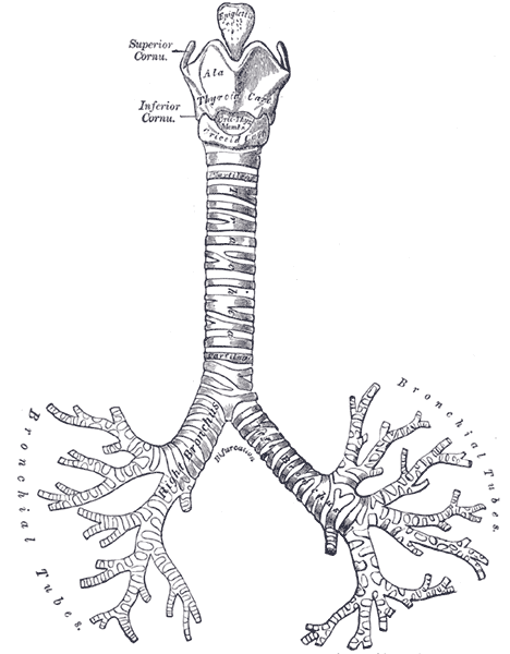
Cartilage-reinforced airway from the larynx (C6) to its bifurcation at the carina (about T4–T5). Its midline position is an important exam and radiographic sign.
Core anatomy
- Location
Neck and superior mediastinum, in the midline anterior to the esophagus.
- Parts
- Begins at the inferior border of the cricoid cartilage (C6)
- 16–20 C-shaped hyaline cartilage rings, open posteriorly, bridged by the trachealis (smooth muscle)
- Carina: internal ridge at the bifurcation into the right and left main bronchi, at about the level of the sternal angle (T4–T5)
- Relations
Anterior (neck): isthmus of the thyroid gland (over rings 2–4), inferior thyroid veins. Anterior (thorax): manubrium, thymus remnant, left brachiocephalic vein, brachiocephalic trunk and left common carotid artery, arch of the aorta. Posterior: Esophagus. Lateral: thyroid lobes and carotid sheaths (neck), the recurrent laryngeal nerves in the tracheoesophageal grooves, the aortic arch (left) and the azygos vein (right).
- Arterial supply
Inferior thyroid arteries (cervical part); bronchial arteries (thoracic part).
- Innervation
Vagus nerve and recurrent laryngeal nerves (sensory and parasympathetic); sympathetic trunk. The carina is highly sensitive (cough reflex).
Forms a boundary of
Found in
Mentioned in
Generated automatically from links in other entries.

Clinical correlations
Tracheal deviation PANCE
- Mechanism / lesion
Pushed away by tension pneumothorax, a large pleural effusion or a mass. Pulled toward by atelectasis or after pneumonectomy, or by fibrosis.
- Classic presentation
Palpable shift of the trachea in the suprasternal notch; mediastinal shift on CXR.
Endotracheal intubation PANCE
- Mechanism / lesion
A tube placed through the glottis into the trachea.
- Classic presentation
The tip should lie a few centimeters above the carina. Too deep usually means a right main bronchus intubation, with left lung collapse and absent left breath sounds. Confirm with end-tidal CO₂, auscultation and chest X-ray. verify
Tracheoesophageal fistula with esophageal atresia
- Mechanism / lesion
Failure of separation of the foregut into the trachea and esophagus. The most common type is proximal esophageal atresia with a distal TE fistula.
- Classic presentation
Newborn with drooling, choking or coughing with feeds, inability to pass an NG tube (it coils on X-ray), and a gas-filled stomach if a distal fistula is present. Associated with VACTERL.
Tracheostomy and tracheal stenosis
- Mechanism / lesion
Surgical airway below the cricoid; stenosis can follow prolonged intubation or tracheostomy.
- Classic presentation
Stenosis causes progressive dyspnea and stridor, sometimes misdiagnosed as asthma.
High-yield PANCE pearls
- Trachea: C6 → T4/T5 (carina at the sternal angle).
- Tension pneumothorax pushes the trachea away; atelectasis pulls it toward.
- Too-deep ETT → right main bronchus; absent breath sounds on the left.
- Newborn with drooling, choking on feeds, NG tube coiling → esophageal atresia ± TE fistula.
Imaging anatomy
- X-ray
The tracheal air column should be midline, projecting over the spinous processes on a non-rotated PA film. Check ETT tip position relative to the carina. A widened carinal angle can suggest left atrial enlargement or subcarinal lymphadenopathy.
- CT
Stenosis, tumors, tracheomalacia (dynamic expiratory CT), and extrinsic compression (goiter, mass).
- Ultrasound
Point-of-care airway ultrasound: the tracheal rings appear as curved hypoechoic arcs anterior to a bright air line. It can confirm ETT placement (a single air–mucosa interface, with the esophagus not distended) and identify the cricothyroid membrane for emergency airway access.
- Key views & landmarks
- PA chest radiograph (tracheal position, ETT tip)
- CT neck/chest for airway narrowing
Physical exam
- Surface landmarks
Palpable in the suprasternal (jugular) notch in the midline. Above it, the cricoid cartilage and the cricothyroid membrane (between the thyroid and cricoid cartilages) are the landmarks for emergency cricothyrotomy.
- Palpation
Place a finger in the suprasternal notch and feel the space on each side of the trachea; asymmetry means deviation.
- Strength / function testing
Auscultate over the trachea for stridor and normal tracheal (loud, harsh) breath sounds.
Self-quiz
After emergency intubation, breath sounds are present on the right and absent on the left. End-tidal CO₂ is detected. What is the most likely problem?
Correct answer: C
A tube advanced too far usually enters the right main bronchus (wider and more vertical), ventilating only the right lung. Withdraw the tube and reconfirm. CO₂ detection rules out esophageal intubation.
Show answer
C
A tube advanced too far usually enters the right main bronchus (wider and more vertical), ventilating only the right lung. Withdraw the tube and reconfirm. CO₂ detection rules out esophageal intubation.
At which vertebral level does the trachea bifurcate?
Correct answer: C
The carina lies at about T4–T5, at the level of the sternal angle. The trachea begins at C6 (cricoid).
Show answer
C
The carina lies at about T4–T5, at the level of the sternal angle. The trachea begins at C6 (cricoid).
A patient's trachea is shifted to the right. Breath sounds and percussion over the left hemithorax are decreased and dull, and the chest X-ray shows a white-out of the left hemithorax. What is the most likely cause?
Correct answer: B
A large left pleural effusion pushes the trachea and mediastinum away (to the right). Left-sided atelectasis or pneumonectomy would pull the trachea toward the white-out.
Show answer
B
A large left pleural effusion pushes the trachea and mediastinum away (to the right). Left-sided atelectasis or pneumonectomy would pull the trachea toward the white-out.
What bridges the posterior gap in the C-shaped tracheal cartilages?
Correct answer: D
The trachea has 16–20 C-shaped hyaline cartilage rings, open posteriorly and bridged by the trachealis smooth muscle, where the trachea lies against the esophagus.
Show answer
D
The trachea has 16–20 C-shaped hyaline cartilage rings, open posteriorly and bridged by the trachealis smooth muscle, where the trachea lies against the esophagus.
A newborn drools, chokes with feeds, and a nasogastric tube coils in the upper chest on X-ray. The stomach is filled with gas. What is the most likely anomaly?
Correct answer: C
The most common type is proximal esophageal atresia with a distal TE fistula: the NG tube coils in the blind pouch, and air reaches the stomach through the fistula. Isolated atresia leaves a gasless abdomen. It is associated with VACTERL.
Show answer
C
The most common type is proximal esophageal atresia with a distal TE fistula: the NG tube coils in the blind pouch, and air reaches the stomach through the fistula. Isolated atresia leaves a gasless abdomen. It is associated with VACTERL.