Lungs
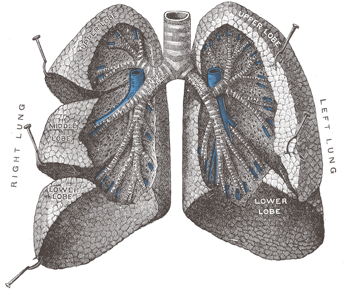
Paired organs of respiration. The right lung has three lobes and the left has two plus the lingula; lobe location drives exam findings and imaging signs.
Core anatomy
- Location
Each lung occupies its pleural cavity on either side of the mediastinum, from the apex (above the clavicle) to the base on the diaphragm.
- Parts
- Right lung: superior, middle and inferior lobes, separated by the oblique and horizontal fissures; 10 bronchopulmonary segments
- Left lung: superior and inferior lobes separated by the oblique fissure; the lingula (part of the superior lobe) is the counterpart of the right middle lobe; cardiac notch; 8–10 segments (some fused)
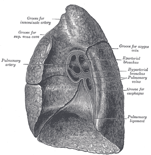
- Hilum (root): main bronchus, pulmonary artery, two pulmonary veins, bronchial vessels, nerves and lymphatics
- Apex, base (diaphragmatic surface), costal and mediastinal surfaces
- Relations
Hilum arrangement: the pulmonary veins are the most anterior and inferior. On the right, the eparterial (superior lobe) bronchus lies superior to the pulmonary artery. On the left, the pulmonary artery is most superior. Mnemonic RALS: the pulmonary artery is Right Anterior, Left Superior to the bronchus. The mediastinal surface bears impressions of the heart, the thoracic aorta (left) and the superior vena cava, azygos vein and esophagus (right).
- Arterial supply
Functional: Pulmonary arteries (deoxygenated blood for gas exchange). Nutritive: bronchial arteries (from the thoracic aorta) supplying the bronchi, connective tissue and visceral pleura.
- Venous drainage
Pulmonary veins (usually two per side) to the left atrium. Bronchial veins to the azygos system (part of the flow joins the pulmonary veins).
- Lymphatic drainage
Pulmonary → bronchopulmonary (hilar) → tracheobronchial (including subcarinal) → bronchomediastinal trunks.
- Innervation
Pulmonary plexuses. Vagus (parasympathetic: bronchoconstriction, secretion) and sympathetic trunk (bronchodilation). Visceral afferents for cough and stretch reflexes. The lungs and visceral pleura are insensitive to pain.
- Notes
Surface markings. Apex: 2–3 cm above the medial third of the clavicle. Oblique fissure: from about the T2–T3 spinous process posteriorly, around the chest to the 6th costal cartilage anteriorly (roughly following the medial border of the abducted scapula). Horizontal fissure (right): along the 4th rib/costal cartilage from the sternum to the mid-axillary line. Inferior border: 6th rib (midclavicular), 8th rib (mid-axillary), 10th rib (posteriorly). So the lower lobes lie mostly posteriorly and the right middle lobe anteriorly. verify


Clinical correlations
Lobar pneumonia and aspiration PANCE
- Mechanism / lesion
Infection of the alveoli. Aspiration favors the right lung because the right main bronchus is wider and more vertical: upright → basal segments of the right lower lobe; supine → the posterior segment of the right upper lobe and the superior segment of the right lower lobe.
- Classic presentation
Fever, productive cough and pleuritic pain. Over the consolidation: dullness, increased fremitus, bronchial breath sounds, egophony, crackles.
Lung cancer by location PANCE
- Mechanism / lesion
Central: squamous cell and small cell carcinoma (associated with smoking). Peripheral: adenocarcinoma (the most common type overall and in non-smokers). Apical (Pancoast) tumors invade the brachial plexus and sympathetic chain.
- Classic presentation
Cough, hemoptysis, weight loss. Local invasion causes hoarseness (left recurrent laryngeal nerve), an elevated hemidiaphragm (phrenic nerve), SVC syndrome, or Horner syndrome (Pancoast). Paraneoplastic syndromes include SIADH and Cushing syndrome (small cell) and hypercalcemia (squamous).
Pulmonary embolism PANCE
- Mechanism / lesion
Thrombus (usually from deep leg veins) lodges in the pulmonary arteries.
- Classic presentation
Sudden dyspnea, pleuritic chest pain, tachycardia, hypoxemia; massive PE causes hypotension and RV strain.
High-yield PANCE pearls
- Right lung: 3 lobes, 2 fissures. Left lung: 2 lobes + lingula, 1 fissure, cardiac notch.
- Aspirated material → right lung (upright: RLL basal; supine: RUL posterior / RLL superior segments).
- Silhouette sign: RML pneumonia obscures the right heart border; lingular pneumonia obscures the left heart border; lower lobe disease obscures the diaphragm.
- Central tumors: squamous and small cell; peripheral: adenocarcinoma.
- Hilum: RALS (pulmonary artery Right Anterior, Left Superior).
Imaging anatomy
- X-ray
PA and lateral chest radiographs. Use the silhouette sign to localize disease: loss of the right heart border = right middle lobe; loss of the left heart border = lingula; loss of the hemidiaphragm = lower lobe. The lateral view shows the fissures and retrocardiac/posterior lower-lobe opacities. Look for air bronchograms (consolidation), volume loss (atelectasis: shift toward), masses, and hyperinflation (COPD: flattened diaphragms).
- CT
CT chest defines nodules, masses, interstitial disease and lymphadenopathy. CT pulmonary angiography (CTPA) is the main test for PE. Low-dose CT is used for lung cancer screening in eligible high-risk smokers.
- Ultrasound
Lung ultrasound shows consolidation ("hepatization", dynamic air bronchograms), interstitial fluid (B-lines, e.g. in pulmonary edema), effusion, and pneumothorax (absent sliding).
- Key views & landmarks
- PA and lateral chest radiograph
- CT chest (CTPA for PE)
Physical exam
- Surface landmarks
Anteriorly you mostly hear the upper lobes (and the right middle lobe below the 4th rib on the right). Posteriorly you mostly hear the lower lobes (below about T3). Laterally you hear upper and lower lobes, plus the middle lobe on the right.
- Palpation
Chest expansion (thumbs at the 10th ribs posteriorly), tactile fremitus.
- Strength / function testing
Percussion (resonant, dull, hyperresonant) and diaphragmatic excursion. Auscultation (vesicular, bronchial and bronchovesicular breath sounds; crackles, wheezes, rhonchi, rubs). Transmitted voice sounds (egophony "E"→"A", bronchophony, whispered pectoriloquy) suggest consolidation.
Self-quiz
An intoxicated man found lying supine develops fever and a foul-smelling productive cough 3 days later. Which lung segment is most likely involved?
Correct answer: C
In the supine position, aspirated material flows to the dependent posterior segment of the right upper lobe and the superior segment of the right lower lobe. Upright aspiration goes to the right lower lobe basal segments.
Show answer
C
In the supine position, aspirated material flows to the dependent posterior segment of the right upper lobe and the superior segment of the right lower lobe. Upright aspiration goes to the right lower lobe basal segments.
On a PA chest radiograph, a patient's right heart border is obscured by an opacity while the right hemidiaphragm is sharply seen. Where is the pneumonia?
Correct answer: B
The right middle lobe lies against the right heart border, so consolidation there obscures it (silhouette sign). Right lower lobe disease obscures the hemidiaphragm instead.
Show answer
B
The right middle lobe lies against the right heart border, so consolidation there obscures it (silhouette sign). Right lower lobe disease obscures the hemidiaphragm instead.
A patient has dullness to percussion, increased tactile fremitus and bronchial breath sounds over the right lower chest posteriorly. What is the most likely diagnosis?
Correct answer: C
Consolidation transmits vibrations and sounds better: dull percussion, increased fremitus, bronchial breath sounds and egophony. An effusion is dull with decreased fremitus.
Show answer
C
Consolidation transmits vibrations and sounds better: dull percussion, increased fremitus, bronchial breath sounds and egophony. An effusion is dull with decreased fremitus.
Where does the horizontal fissure of the right lung lie anteriorly?
Correct answer: B
The horizontal fissure follows the 4th rib and costal cartilage from the sternum to about the mid-axillary line, where it meets the oblique fissure.
Show answer
B
The horizontal fissure follows the 4th rib and costal cartilage from the sternum to about the mid-axillary line, where it meets the oblique fissure.
Which part of the left lung corresponds to the right middle lobe?
Correct answer: E
The lingula of the left upper lobe is the counterpart of the right middle lobe. Lingular pneumonia obscures the left heart border, just as right middle lobe pneumonia obscures the right heart border (silhouette sign).
Show answer
E
The lingula of the left upper lobe is the counterpart of the right middle lobe. Lingular pneumonia obscures the left heart border, just as right middle lobe pneumonia obscures the right heart border (silhouette sign).