Perineal pouches
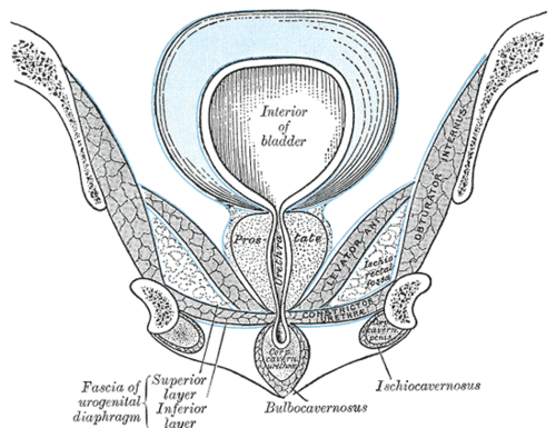
Two fascial compartments of the urogenital triangle, separated by the perineal membrane. Their fascial attachments determine where urine spreads after urethral injury and how Fournier gangrene tracks.
Core anatomy
- Location
In the urogenital triangle of the perineum, between the ischiopubic rami.
- Boundaries
- superficial pouch — inferior
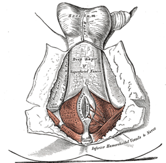
- Membranous layer of the superficial perineal fascia (Colles fascia), continuous with the dartos fascia and Scarpa fascia of the abdominal wall
- superficial pouch — superior
- Perineal membrane
- deep pouch — inferior
- Perineal membrane
- deep pouch — superior
- Inferior fascia of the pelvic diaphragm (the deep pouch is now regarded as open superiorly toward the pelvis)
- lateral
- Ischiopubic rami (where Colles fascia and the perineal membrane attach)
- posterior
- Fusion of Colles fascia with the posterior border of the perineal membrane at the perineal body
- Contents
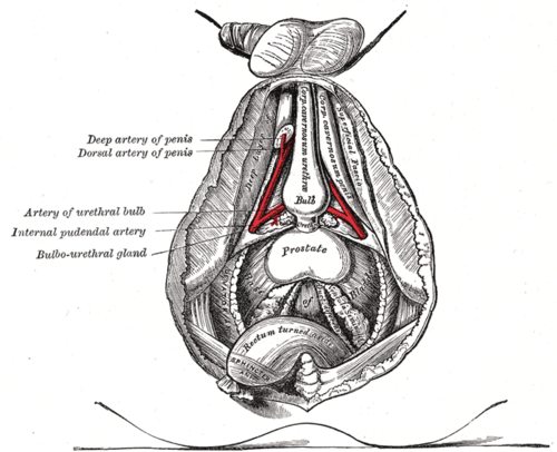
- Superficial pouch (male): root of the penis (bulb and crura), bulbospongiosus, ischiocavernosus, superficial transverse perineal muscles, the proximal spongy urethra, and branches of the internal pudendal vessels and pudendal nerve
- Superficial pouch (female): clitoris (crura), bulbs of the vestibule, greater vestibular (Bartholin) glands, the same muscles (see vulva)
- Deep pouch (male): the intermediate (membranous) urethra, the external urethral sphincter, deep transverse perineal muscle, bulbourethral (Cowper) glands, and the dorsal vessels and nerves of the penis
- Deep pouch (female): the proximal urethra, the external urethral sphincter with compressor urethrae and urethrovaginal sphincter, and the dorsal vessels and nerves of the clitoris
- Communications
The superficial pouch is open anteriorly to the space beneath the membranous (Scarpa) layer of the anterior abdominal wall and the dartos of the scrotum and penis. It is closed laterally (ischiopubic rami), posteriorly (perineal body) and at the thighs (Scarpa/Colles fascia fuses with the fascia lata just below the inguinal ligament).
Swipe sideways to see the whole diagram →



Clinical correlations
Extravasation of urine after bulbar urethral rupture PANCE
- Mechanism / lesion
A straddle injury tears the spongy (bulbar) urethra and Buck fascia in the superficial pouch.
- Classic presentation
Urine and blood spread into the scrotum, penis and anterior abdominal wall deep to Scarpa fascia, but not into the thighs (fusion with the fascia lata) or the anal triangle (fusion at the perineal body). Classic "butterfly" perineal bruising outlines Colles fascia.
Membranous urethral injury PANCE
- Mechanism / lesion
Pelvic fracture shears the membranous urethra in the deep pouch.
- Classic presentation
Extravasation into the deep pouch and the retropubic (extraperitoneal) space around the bladder, with blood at the meatus and inability to void. Retrograde urethrogram before catheterization.
Spread of Fournier gangrene
- Mechanism / lesion
Necrotizing infection tracks along Colles, dartos and Scarpa fascia.
- Classic presentation
It can spread rapidly from the perineum to the scrotum, penis and anterior abdominal wall; the testes are usually spared because they have their own blood supply. See perineum.
High-yield PANCE pearls
- Bulbar (straddle) urethral injury → urine into scrotum, penis, abdominal wall — not thighs.
- Membranous urethral injury (pelvic fracture) → deep pouch and retropubic space.
- Colles fascia = perineal continuation of Scarpa fascia.
- Bulbourethral glands are in the deep pouch; Bartholin glands in the superficial pouch.
Imaging anatomy
- X-ray
Retrograde urethrogram localizes the urethral tear and the pattern of extravasation.
- CT
CT shows perineal hematoma, fluid and gas along fascial planes.
- MRI
MRI defines perineal anatomy for urethral stricture planning and after trauma.
Physical exam
- Surface landmarks
The perineal membrane spans the pubic arch between the ischiopubic rami; the bulb of the penis lies in the midline beneath bulbospongiosus.
- Palpation
Inspect for a butterfly-shaped perineal hematoma, scrotal and penile swelling and blood at the meatus; check the distribution of swelling (spared thighs point to the superficial pouch).
Self-quiz
The external urethral sphincter and bulbourethral glands in a male are located in which compartment?
Correct answer: B
The deep perineal pouch contains the membranous urethra, the external urethral sphincter and the bulbourethral (Cowper) glands. The root of the penis and its muscles are in the superficial pouch.
Show answer
B
The deep perineal pouch contains the membranous urethra, the external urethral sphincter and the bulbourethral (Cowper) glands. The root of the penis and its muscles are in the superficial pouch.
Why does urine from a ruptured bulbar urethra not spread into the thighs?
Correct answer: B
The membranous layer of superficial fascia (Scarpa in the abdomen, Colles in the perineum) fuses with the fascia lata just below the inguinal ligament, closing off the thigh; it also fuses with the perineal membrane posteriorly, closing off the anal triangle.
Show answer
B
The membranous layer of superficial fascia (Scarpa in the abdomen, Colles in the perineum) fuses with the fascia lata just below the inguinal ligament, closing off the thigh; it also fuses with the perineal membrane posteriorly, closing off the anal triangle.
In a woman, the greater vestibular (Bartholin) glands lie in which compartment?
Correct answer: B
The superficial perineal pouch in women contains the crura of the clitoris, the bulbs of the vestibule, the greater vestibular (Bartholin) glands and the superficial perineal muscles. In men, the bulbourethral (Cowper) glands are in the deep pouch.
Show answer
B
The superficial perineal pouch in women contains the crura of the clitoris, the bulbs of the vestibule, the greater vestibular (Bartholin) glands and the superficial perineal muscles. In men, the bulbourethral (Cowper) glands are in the deep pouch.
Colles fascia of the perineum is continuous with which layer of the anterior abdominal wall?
Correct answer: E
Colles fascia (the membranous layer of the superficial perineal fascia) forms the floor of the superficial pouch and is continuous with the dartos fascia and the membranous (Scarpa) layer of the abdominal wall. This is why urine from a bulbar urethral rupture, or Fournier gangrene, can spread up onto the abdominal wall.
Show answer
E
Colles fascia (the membranous layer of the superficial perineal fascia) forms the floor of the superficial pouch and is continuous with the dartos fascia and the membranous (Scarpa) layer of the abdominal wall. This is why urine from a bulbar urethral rupture, or Fournier gangrene, can spread up onto the abdominal wall.
A pelvic fracture tears the membranous urethra. Where does the urine extravasate?
Correct answer: D
The membranous urethra lies in the deep pouch, so a pelvic-fracture injury leaks into the deep pouch and the retropubic space around the bladder. Spread into the scrotum, penis and abdominal wall is typical of a bulbar (straddle) injury in the superficial pouch. Obtain a retrograde urethrogram before catheterization.
Show answer
D
The membranous urethra lies in the deep pouch, so a pelvic-fracture injury leaks into the deep pouch and the retropubic space around the bladder. Spread into the scrotum, penis and abdominal wall is typical of a bulbar (straddle) injury in the superficial pouch. Obtain a retrograde urethrogram before catheterization.
Flashcards
See also
- Urethra
- Perineum
- Penis
- Vulva
- Blood at the urethral meatus (clinical case)