Diaphragm
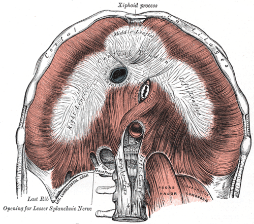
Dome-shaped musculotendinous partition between the thorax and abdomen. It is the main muscle of inspiration and is supplied by the phrenic nerves (C3–C5).
Core anatomy
- Location
Between the thoracic and abdominal cavities. The right dome sits slightly higher than the left (liver below).
- Group
Muscles of respiration.
- Origin
Sternal part: posterior xiphoid process. Costal part: internal surfaces of the lower six costal cartilages and adjacent ribs. Lumbar part: medial and lateral arcuate ligaments and the crura; the right crus arises from the bodies of L1–L3, the left from L1–L2.
- Insertion
Central tendon (converging from all parts).
- Innervation
Motor: Phrenic nerve (C3, C4, C5), the sole motor supply. Sensory: phrenic nerve to the central part; the lower intercostal and subcostal nerves to the peripheral costal part.
- Blood supply
Pericardiacophrenic and musculophrenic arteries (from the internal thoracic artery), superior phrenic arteries (thoracic aorta), and inferior phrenic arteries (abdominal aorta).
- Action
- Principal muscle of inspiration. Contraction flattens and lowers the domes, increasing thoracic volume
- Increases intra-abdominal pressure (with the abdominal wall) for coughing, vomiting, defecation and lifting
- Relations
Openings (approximate vertebral levels): caval opening, T8, in the central tendon (inferior vena cava, branches of the right phrenic nerve); esophageal hiatus, T10, in the right crus (esophagus, anterior and posterior vagal trunks, esophageal branches of the left gastric vessels); aortic hiatus, T12, behind the median arcuate ligament (aorta, thoracic duct, and often the azygos vein). Mnemonic: "I 8 10 Eggs At 12" (IVC T8, esophagus T10, aorta T12).
Forms a boundary of
Mentioned in
Generated automatically from links in other entries.

Clinical correlations
Hiatal hernia PANCE
- Mechanism / lesion
Herniation of the stomach through the esophageal hiatus. In a sliding hernia (type I, the most common) the gastroesophageal junction moves above the diaphragm. In a paraesophageal hernia the fundus herniates beside a GE junction that stays in place.
- Classic presentation
Sliding: GERD symptoms (heartburn, regurgitation). Paraesophageal: often asymptomatic, but risk of incarceration or volvulus (chest pain, retching, inability to swallow).
Congenital diaphragmatic hernia PANCE
- Mechanism / lesion
Defective closure of the pleuroperitoneal membrane, most often posterolateral and left-sided (Bochdalek). Abdominal viscera in the thorax impair lung development (pulmonary hypoplasia).
- Classic presentation
Newborn with respiratory distress, a scaphoid abdomen, bowel sounds in the chest, and shift of heart sounds. Often detected on prenatal ultrasound.
Diaphragmatic paralysis
- Mechanism / lesion
Phrenic nerve injury (cardiac or thoracic surgery, tumor invasion, interscalene block, trauma, cervical spinal cord injury at C3–C5).
- Classic presentation
Unilateral: often asymptomatic or exertional dyspnea, with an elevated hemidiaphragm on CXR. Bilateral: orthopnea and paradoxical inward movement of the abdomen on inspiration.
Traumatic diaphragmatic rupture
- Mechanism / lesion
Blunt or penetrating thoracoabdominal trauma; blunt ruptures are more often on the left (the liver protects the right).
- Classic presentation
Abdominal contents in the chest on imaging (e.g. an NG tube curling in the left chest). Easily missed initially.
High-yield PANCE pearls
- Openings: IVC T8, esophagus T10, aorta T12 ("I 8 10 Eggs At 12").
- "C3, 4, 5 keeps the diaphragm alive" (phrenic nerve).
- Diaphragmatic irritation → referred shoulder pain (C3–C5), e.g. Kehr sign with splenic rupture.
- Newborn with respiratory distress + scaphoid abdomen → congenital diaphragmatic hernia (left Bochdalek).
- Free air under the diaphragm on an upright CXR → perforated viscus.
Imaging anatomy
- X-ray
Upright CXR: the domes project at about the 5th–6th anterior ribs. The right dome is usually higher. Free intraperitoneal air appears as a lucent crescent under the diaphragm. An elevated hemidiaphragm suggests paralysis, eventration, subpulmonic effusion or volume loss. Blunting of the costophrenic angle suggests effusion. verify
- CT
Multiplanar CT shows hernias, traumatic rupture ("collar sign" where herniated viscus is constricted by the torn diaphragm) and masses.
- MRI
Occasionally used to confirm diaphragmatic defects when CT is equivocal.
- Ultrasound
M-mode ultrasound measures diaphragmatic excursion and thickening, identifying paralysis at the bedside. It also shows effusions above the diaphragm and free fluid below it.
- Key views & landmarks
- Upright PA and lateral CXR
- Fluoroscopic sniff test: a paralyzed hemidiaphragm moves paradoxically upward on a sharp sniff
Physical exam
- Surface landmarks
The domes rise to about the 5th intercostal space in expiration; excursion is roughly 3–5 cm in adults. verify
- Palpation
Not directly palpable.
- Strength / function testing
Percussion of diaphragmatic excursion posteriorly: find the level of dullness on full expiration and full inspiration. Reduced excursion suggests paralysis, effusion or hyperinflation. Observe for paradoxical abdominal movement when supine.
Self-quiz
At approximately which vertebral level does the esophagus pass through the diaphragm?
Correct answer: B
The esophageal hiatus is at T10, with the vagal trunks. The IVC passes at T8 through the central tendon; the aorta at T12 behind the median arcuate ligament.
Show answer
B
The esophageal hiatus is at T10, with the vagal trunks. The IVC passes at T8 through the central tendon; the aorta at T12 behind the median arcuate ligament.
A term newborn has respiratory distress immediately after birth. The abdomen is scaphoid and bowel sounds are heard over the left chest. What is the most likely diagnosis?
Correct answer: B
A Bochdalek (posterolateral, usually left) congenital diaphragmatic hernia puts bowel in the chest, leaving a scaphoid abdomen, and causes pulmonary hypoplasia.
Show answer
B
A Bochdalek (posterolateral, usually left) congenital diaphragmatic hernia puts bowel in the chest, leaving a scaphoid abdomen, and causes pulmonary hypoplasia.
A patient with splenic rupture reports pain at the tip of the left shoulder. Which nerve carries the referred pain?
Correct answer: B
Blood irritating the diaphragm stimulates phrenic nerve (C3–C5) sensory fibers. The pain is referred to the shoulder, which shares the C3–C5 dermatomes (Kehr sign).
Show answer
B
Blood irritating the diaphragm stimulates phrenic nerve (C3–C5) sensory fibers. The pain is referred to the shoulder, which shares the C3–C5 dermatomes (Kehr sign).
Which structure passes through the aortic hiatus of the diaphragm with the aorta?
Correct answer: B
The aortic hiatus (T12) transmits the aorta, the thoracic duct and often the azygos vein. The caval opening (T8) transmits the IVC and branches of the right phrenic nerve; the esophageal hiatus (T10) transmits the esophagus, the vagal trunks and esophageal branches of the left gastric vessels.
Show answer
B
The aortic hiatus (T12) transmits the aorta, the thoracic duct and often the azygos vein. The caval opening (T8) transmits the IVC and branches of the right phrenic nerve; the esophageal hiatus (T10) transmits the esophagus, the vagal trunks and esophageal branches of the left gastric vessels.
After blunt abdominal trauma, a chest radiograph shows a nasogastric tube curling up into the left hemithorax. Why are traumatic diaphragmatic ruptures more often left-sided?
Correct answer: E
Blunt diaphragmatic ruptures are more often left-sided because the liver buffers the right side. Abdominal contents (e.g. the stomach with an NG tube) in the chest are the clue, and the injury is easily missed.
Show answer
E
Blunt diaphragmatic ruptures are more often left-sided because the liver buffers the right side. Abdominal contents (e.g. the stomach with an NG tube) in the chest are the clue, and the injury is easily missed.