Stomach
J-shaped dilated foregut organ in the upper abdomen, supplied by all three branches of the celiac trunk. Peptic ulcers, gastric cancer and pyloric stenosis are its key clinical problems.
Core anatomy
- Location
Mainly in the left upper quadrant, epigastric and umbilical regions. Position varies with body habitus and filling.
- Parts
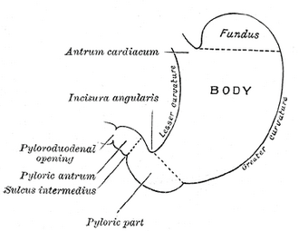
- Cardia (around the gastroesophageal junction), fundus (above the cardiac notch), body, pyloric antrum, pyloric canal, pylorus (pyloric sphincter, at about the transpyloric plane, L1)
- Lesser curvature (with the angular incisure) and greater curvature
- Gastric folds (rugae); physiological lower esophageal sphincter at the GE junction
- Relations
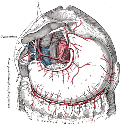
Anterior: left lobe of the liver, diaphragm, anterior abdominal wall. Posterior (the "stomach bed"), separated by the lesser sac: Pancreas, left kidney and suprarenal gland, spleen, splenic artery, transverse mesocolon. The greater omentum hangs from the greater curvature; the lesser omentum runs from the lesser curvature to the liver.
- Arterial supply
All from the celiac trunk. Lesser curvature: left gastric artery (direct branch of the celiac trunk) and right gastric artery (usually from the proper or common hepatic artery). Greater curvature: right gastro-omental (from the gastroduodenal) and left gastro-omental (from the splenic). Fundus: short gastric arteries (from the splenic), plus the posterior gastric artery.
- Venous drainage
Gastric veins to the hepatic portal vein system. The left gastric vein anastomoses with esophageal veins (esophageal varices in portal hypertension).
- Lymphatic drainage
Gastric and gastro-omental nodes → celiac nodes. Spread can reach the left supraclavicular node (Virchow).
- Innervation
Parasympathetic: anterior and posterior vagal trunks (acid secretion, motility). Sympathetic: T6–T9 via the greater splanchnic nerves and celiac plexus. Visceral pain refers to the epigastrium (foregut).
Mentioned in
Generated automatically from links in other entries.


Clinical correlations
Peptic ulcer disease PANCE
- Mechanism / lesion
Mucosal injury, most often from Helicobacter pylori and NSAIDs (also Zollinger–Ellison syndrome, smoking). Duodenal ulcers are more common than gastric ulcers.
- Classic presentation
Burning epigastric pain. Classically, gastric ulcer pain worsens with meals and duodenal ulcer pain improves with meals (and occurs at night). Complications: bleeding (a lesser-curvature ulcer can erode the left gastric artery), perforation (free air), and gastric outlet obstruction. Gastric ulcers need biopsy to exclude cancer.
Gastric adenocarcinoma PANCE
- Mechanism / lesion
Risk factors include H. pylori, smoked/salted foods, pernicious anemia/atrophic gastritis, and prior partial gastrectomy.
- Classic presentation
Weight loss, early satiety, epigastric pain, anemia. Signs of spread: Virchow node (left supraclavicular), Sister Mary Joseph nodule (periumbilical), Krukenberg tumor (ovary), Blumer shelf (rectouterine pouch).
Hypertrophic pyloric stenosis PANCE
- Mechanism / lesion
Hypertrophy of the pyloric muscle in early infancy (typically 3–6 weeks old, more common in first-born males).
- Classic presentation
Projectile, non-bilious vomiting after feeds with a hungry infant, a palpable "olive" in the epigastrium, visible peristalsis, and hypochloremic, hypokalemic metabolic alkalosis. Treated with pyloromyotomy after correcting fluids and electrolytes.
High-yield PANCE pearls
- Gastric ulcer: pain worse with food. Duodenal ulcer: pain better with food; more common.
- PUD causes: H. pylori and NSAIDs; test and treat H. pylori.
- Pyloric stenosis: 3–6-week-old, projectile non-bilious vomiting, olive, hypochloremic hypokalemic metabolic alkalosis; ultrasound to diagnose.
- Gastric cancer signs: Virchow node, Sister Mary Joseph nodule, Krukenberg tumor.
- Stomach blood supply is entirely from the celiac trunk.
Imaging anatomy
- X-ray
Upright CXR for free air after perforation. Upper GI contrast series shows ulcers, masses, outlet obstruction, and in infants the "string sign" of pyloric stenosis (ultrasound is preferred).
- CT
Gastric wall thickening or mass, perforation, cancer staging.
- Ultrasound
First-line for pyloric stenosis: a thickened, elongated pyloric channel (quoted thresholds around 3–4 mm muscle thickness and about 15 mm or more channel length). verify
- Key views & landmarks
- Upper endoscopy (EGD) with biopsy: the diagnostic procedure for ulcers and cancer
- Pyloric ultrasound in infants
Physical exam
- Surface landmarks
The stomach projects to the left upper quadrant and epigastrium. The pylorus lies near the transpyloric plane (L1), just right of the midline.
- Palpation
Epigastric tenderness in PUD. In infants, palpate for the olive in the epigastrium after the stomach is emptied.
- Strength / function testing
A succussion splash (sloshing heard with a stethoscope while rocking the patient) more than a few hours after a meal suggests gastric outlet obstruction.
Self-quiz
A 4-week-old first-born boy has projectile non-bilious vomiting after every feed and is eager to feed again. Which metabolic abnormality is expected?
Correct answer: B
Hypertrophic pyloric stenosis causes loss of gastric HCl through vomiting, producing a hypochloremic, hypokalemic metabolic alkalosis. Ultrasound confirms the diagnosis.
Show answer
B
Hypertrophic pyloric stenosis causes loss of gastric HCl through vomiting, producing a hypochloremic, hypokalemic metabolic alkalosis. Ultrasound confirms the diagnosis.
A patient with a bleeding ulcer on the lesser curvature of the stomach is most likely bleeding from which artery?
Correct answer: B
Lesser-curvature ulcers typically erode the left gastric artery. Posterior duodenal ulcers erode the gastroduodenal artery.
Show answer
B
Lesser-curvature ulcers typically erode the left gastric artery. Posterior duodenal ulcers erode the gastroduodenal artery.
A 45-year-old man has epigastric pain that improves after eating and wakes him at night. Which is the most likely cause?
Correct answer: B
Duodenal ulcer pain classically improves with meals (food buffers acid) and occurs at night. Gastric ulcer pain typically worsens with eating.
Show answer
B
Duodenal ulcer pain classically improves with meals (food buffers acid) and occurs at night. Gastric ulcer pain typically worsens with eating.
Which branch of the celiac trunk supplies the lesser curvature of the stomach directly?
Correct answer: C
The left gastric artery arises directly from the celiac trunk and runs along the lesser curvature, anastomosing with the right gastric artery.
Show answer
C
The left gastric artery arises directly from the celiac trunk and runs along the lesser curvature, anastomosing with the right gastric artery.
A patient with gastric adenocarcinoma has a hard nodule at the umbilicus. What is this called?
Correct answer: C
A Sister Mary Joseph nodule is a periumbilical metastasis. Other signs of spread: Virchow node (left supraclavicular), Krukenberg tumor (ovary) and Blumer shelf (rectouterine pouch).
Show answer
C
A Sister Mary Joseph nodule is a periumbilical metastasis. Other signs of spread: Virchow node (left supraclavicular), Krukenberg tumor (ovary) and Blumer shelf (rectouterine pouch).
Flashcards
See also
- Duodenum
- Celiac trunk
- Greater omentum
- Foregut, midgut and hindgut (comparison table)