Vertebral ligaments
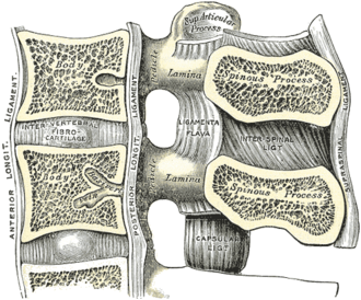
Ligaments that bind the vertebrae. The longitudinal ligaments run along the bodies and the ligamentum flavum joins the laminae; these are the layers a needle passes through during lumbar puncture and epidural anesthesia.
Core anatomy
- Location
Along the full length of the vertebral column.
- Attachments
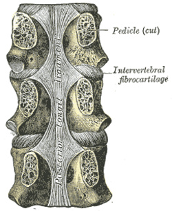
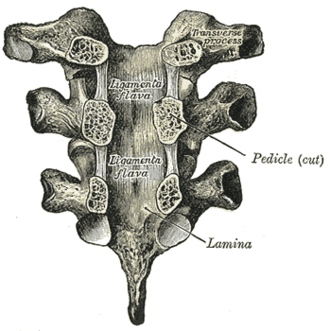
Anterior longitudinal ligament (ALL): broad band on the anterior surfaces of the vertebral bodies and discs, from the occiput and atlas to the sacrum. Posterior longitudinal ligament (PLL): narrower band on the posterior surfaces of the bodies within the canal (continuous above as the tectorial membrane). Ligamentum flavum: joins the laminae of adjacent vertebrae (yellow, elastic). Interspinous ligaments: between spinous processes. Supraspinous ligament: along the tips of the spinous processes, expanding in the neck as the nuchal ligament (ligamentum nuchae). Intertransverse ligaments: between transverse processes.
- Function
The ALL resists hyperextension; the PLL and ligamentum flavum resist hyperflexion (the flavum's elasticity helps restore the upright posture). The PLL's narrowness in the lumbar region leaves the posterolateral disc unsupported.
- Relations
The ligamentum flavum forms the posterior boundary of the epidural space and canal; hypertrophy narrows the canal.
Swipe sideways to see the whole diagram →



Clinical correlations
Layers traversed by a lumbar puncture or epidural needle PANCE
- Mechanism / lesion
Midline approach between spinous processes.
- Classic presentation
Skin → subcutaneous tissue → supraspinous ligament → interspinous ligament → ligamentum flavum (a distinct resistance, then "loss of resistance" on entering the epidural space, where an epidural needle stops) → dura mater → arachnoid mater → subarachnoid space (CSF, for spinal puncture).
Ligamentum flavum hypertrophy and OPLL
- Mechanism / lesion
Ligamentum flavum thickening (with facet and disc degeneration) narrows the canal in lumbar and cervical stenosis. Ossification of the PLL (OPLL) is a cause of cervical myelopathy, more common in East Asian populations.
- Classic presentation
Neurogenic claudication (lumbar) or myelopathy (cervical).
Diffuse idiopathic skeletal hyperostosis (DISH)
- Mechanism / lesion
Flowing ossification along the anterior longitudinal ligament over at least four contiguous vertebrae, typically in older adults (associated with diabetes and obesity); disc spaces and SI joints are preserved.
- Classic presentation
Stiffness, mild pain, dysphagia from cervical osteophytes. Distinguished from ankylosing spondylitis radiographically.
High-yield PANCE pearls
- LP/epidural needle path: supraspinous → interspinous → ligamentum flavum → epidural space → dura → arachnoid → CSF.
- ALL resists hyperextension; PLL resists hyperflexion and is narrow in the lumbar spine.
- Ligamentum flavum hypertrophy → spinal stenosis.
Imaging anatomy
- X-ray
DISH shows flowing anterior ossification with preserved disc spaces. OPLL may be visible on a lateral cervical film.
- CT
Best for ossified ligaments (OPLL, DISH).
- MRI
Ligament injury after trauma (disruption of the PLL or interspinous ligaments suggests instability); ligamentum flavum thickness.
- Ultrasound
Pre-procedure ultrasound of the lumbar spine can identify the midline, interspaces and the ligamentum flavum–dura complex, especially in obese patients.
- Key views & landmarks
- CT (ossification); MRI (ligamentous injury)
Physical exam
- Surface landmarks
The nuchal ligament is palpable in the posterior neck midline; interspinous spaces open with lumbar flexion (fetal position) for LP.
- Strength / function testing
During LP or epidural placement, note the increased resistance of the ligamentum flavum and the loss of resistance on entering the epidural space.
Self-quiz
During an epidural, which structure provides the last firm resistance before the needle enters the epidural space?
Correct answer: C
The ligamentum flavum gives a characteristic firm resistance; the sudden "loss of resistance" marks entry into the epidural space. The dura is deeper (spinal puncture).
Show answer
C
The ligamentum flavum gives a characteristic firm resistance; the sudden "loss of resistance" marks entry into the epidural space. The dura is deeper (spinal puncture).
Which ligament primarily resists hyperextension of the spine?
Correct answer: D
The anterior longitudinal ligament on the front of the vertebral bodies resists hyperextension.
Show answer
D
The anterior longitudinal ligament on the front of the vertebral bodies resists hyperextension.
Which structure connects the laminae of adjacent vertebrae?
Correct answer: C
The elastic ligamentum flavum joins adjacent laminae, forming part of the posterior wall of the canal.
Show answer
C
The elastic ligamentum flavum joins adjacent laminae, forming part of the posterior wall of the canal.
A 70-year-old man with diabetes has spinal stiffness. Radiographs show flowing ossification along the front of at least four contiguous vertebrae, with preserved disc spaces and normal sacroiliac joints. Which ligament is ossified?
Correct answer: B
DISH is flowing ossification of the anterior longitudinal ligament over at least four contiguous vertebrae, associated with diabetes and obesity, with preserved discs and SI joints (unlike ankylosing spondylitis). Ossification of the PLL (OPLL) causes cervical myelopathy.
Show answer
B
DISH is flowing ossification of the anterior longitudinal ligament over at least four contiguous vertebrae, associated with diabetes and obesity, with preserved discs and SI joints (unlike ankylosing spondylitis). Ossification of the PLL (OPLL) causes cervical myelopathy.
The supraspinous ligament expands in the neck as which structure?
Correct answer: C
The supraspinous ligament runs along the tips of the spinous processes and expands in the neck as the nuchal ligament. The PLL continues above as the tectorial membrane.
Show answer
C
The supraspinous ligament runs along the tips of the spinous processes and expands in the neck as the nuchal ligament. The PLL continues above as the tectorial membrane.