Radial nerve
Largest branch of the brachial plexus (posterior cord). It supplies all extensor muscles of the arm and forearm and winds around the humerus in the radial groove.
Core anatomy
- Location
Posterior compartment of the arm → lateral elbow → posterior compartment of the forearm (via its deep branch) and dorsum of the hand (via its superficial branch).
- Root values
C5, C6, C7, C8, T1
- Origin
Terminal branch of the posterior cord of the brachial plexus (its largest branch).
- Course
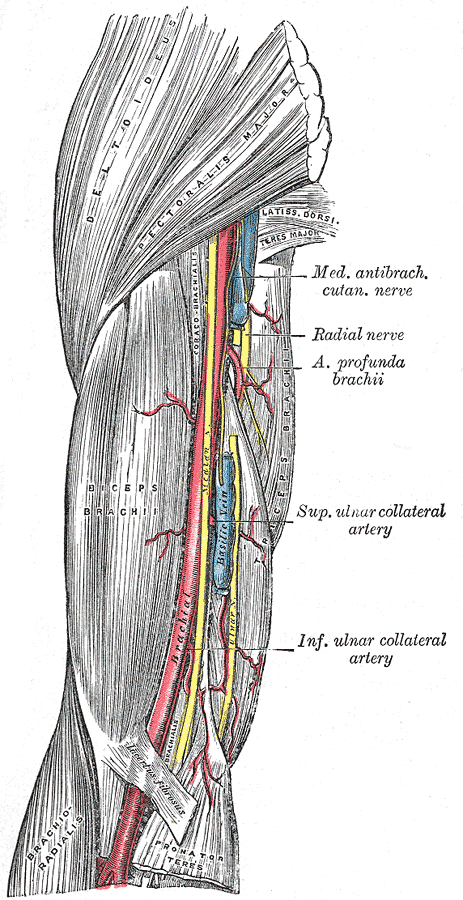
Leaves the axilla posterior to the axillary artery and passes through the triangular interval (below teres major, between the long head of triceps brachii and the humerus) with the profunda brachii (deep brachial) artery. It then spirals around the posterior humerus in the radial groove (spiral groove) between the lateral and medial heads of triceps. On the lateral side of the arm it pierces the lateral intermuscular septum and descends anterior to the lateral epicondyle between brachialis and brachioradialis. In the cubital fossa it divides into:
- Deep branch: motor. It passes between the two layers of supinator (entering under its proximal edge, the arcade of Frohse) and continues as the posterior interosseous nerve (PIN).
- Superficial branch: sensory. It runs under brachioradialis and emerges in the distal forearm to supply the dorsum of the hand.
- Branches
- Posterior cutaneous nerve of the arm; inferior lateral cutaneous nerve of the arm; posterior cutaneous nerve of the forearm
- Muscular branches to triceps and anconeus (mostly arising proximal to the radial groove)
- Muscular branches to brachioradialis and extensor carpi radialis longus (above the elbow)
- Deep branch / posterior interosseous nerve: extensor carpi radialis brevis, supinator, and the posterior forearm extensors
- Superficial branch: sensory to the lateral dorsum of the hand
- Motor
Arm: Triceps brachii, anconeus. Forearm (radial nerve proper): brachioradialis, extensor carpi radialis longus. Deep branch/PIN: extensor carpi radialis brevis, supinator, extensor digitorum, extensor digiti minimi, extensor carpi ulnaris, abductor pollicis longus, extensor pollicis brevis, extensor pollicis longus, extensor indicis. verify
- Sensory
Posterior arm and forearm, the inferolateral arm, and the lateral two-thirds of the dorsum of the hand with the dorsum of the lateral 3½ digits proximal to the nail beds. The most reliable test site is the dorsal first web space.
Muscles innervated
- Triceps brachii — radial nerve (C6, C7, C8)
Other structures innervated
Found in
Mentioned in
Generated automatically from links in other entries.
Swipe sideways to see the whole diagram →

Clinical correlations
Midshaft humerus fracture (radial groove) PANCE
- Mechanism / lesion
Fracture of the humeral shaft injures the nerve where it lies against the bone in the radial groove. A spiral fracture of the distal third of the shaft (Holstein–Lewis fracture) is classically associated with radial nerve palsy.
- Classic presentation
Wrist drop: the wrist and the MCP joints can't be extended. Sensory loss on the dorsum of the hand (first web space). Triceps is usually spared because its branches arise proximal to the groove.
Compression in the axilla or arm ("crutch palsy", "Saturday night palsy") PANCE
- Mechanism / lesion
Axillary compression from improperly used crutches, or compression against the humerus when the arm hangs over a chair back during deep sleep or intoxication.
- Classic presentation
Wrist drop and dorsal hand numbness. With axillary compression, triceps is also weak and the triceps reflex is reduced, because the lesion is proximal to the triceps branches.
Posterior interosseous nerve syndrome
- Mechanism / lesion
Compression of the deep branch at the arcade of Frohse/supinator (repetitive pronation–supination, masses such as lipoma or ganglion, radial head fracture or dislocation).
- Classic presentation
Finger drop without wrist drop: MCP extension of the fingers and thumb is weak. The wrist extends with radial deviation because ECRL (supplied above the elbow) is preserved while ECU is paralyzed. No sensory loss.
Superficial radial neuropathy (Wartenberg syndrome)
- Mechanism / lesion
Compression of the superficial branch at the distal forearm by tight watchbands, handcuffs or casts.
- Classic presentation
Numbness or paresthesias over the dorsoradial hand and thumb with no weakness.
High-yield PANCE pearls
- Midshaft humerus fracture → radial nerve → wrist drop; triceps usually spared.
- Crutch palsy (axilla) → wrist drop plus triceps weakness.
- PIN syndrome = finger drop, wrist extends with radial deviation, no sensory loss.
- Radial nerve territory in the hand: dorsal first web space.
- Reflexes carried by the radial nerve: triceps (C7) and brachioradialis (C6).
Imaging anatomy
- X-ray
The nerve is not visible. AP and lateral humerus radiographs identify shaft fractures, particularly distal-third spiral fractures, which carry the highest association with radial nerve palsy.
- MRI
Denervation edema of supinator and the forearm extensors in PIN syndrome. MRI can show a compressive mass (ganglion, lipoma) at the arcade of Frohse.
- Ultrasound
High-resolution ultrasound can follow the nerve along the spiral groove after humeral fracture to check continuity or entrapment by fragments, and can image the deep branch entering supinator.
- Key views & landmarks
- Humerus AP and lateral (include shoulder and elbow)
- Ultrasound along the posterolateral humerus, spiral groove
Physical exam
- Surface landmarks
Crosses the posterior humerus roughly at mid-shaft. The superficial branch can be rolled over the distal radius just proximal to the anatomical snuffbox.
- Strength / function testing
Wrist extension, finger extension at the MCP joints, and thumb extension against resistance. Triceps strength (elbow extension). Triceps (C7) and brachioradialis (C6) reflexes. Sensation over the dorsal first web space. Localize the lesion: triceps weak → axilla; triceps spared with wrist drop → radial groove; wrist extends with radial deviation but fingers drop → PIN.
Self-quiz
A 34-year-old man sustains a midshaft fracture of the right humerus. He cannot extend his wrist or fingers and has decreased sensation over the dorsum of the first web space. Elbow extension is normal. Where is the nerve injured?
Correct answer: B
Wrist drop with dorsal-hand sensory loss but preserved triceps localizes to the radial groove, because the triceps branches arise proximally. An axillary lesion would weaken triceps too. A lesion at the arcade of Frohse (PIN) causes no sensory loss and spares wrist extension through ECRL.
Show answer
B
Wrist drop with dorsal-hand sensory loss but preserved triceps localizes to the radial groove, because the triceps branches arise proximally. An axillary lesion would weaken triceps too. A lesion at the arcade of Frohse (PIN) causes no sensory loss and spares wrist extension through ECRL.
A 45-year-old carpenter cannot extend his fingers at the MCP joints. When he extends the wrist, it deviates radially. Sensation is normal throughout the hand. Which nerve is most likely compressed?
Correct answer: D
PIN syndrome: the deep branch is compressed at the supinator. ECRL (supplied before the division) still extends the wrist, but ECU is paralyzed, so the wrist deviates radially. The PIN carries no cutaneous fibres, so sensation is normal.
Show answer
D
PIN syndrome: the deep branch is compressed at the supinator. ECRL (supplied before the division) still extends the wrist, but ECU is paralyzed, so the wrist deviates radially. The PIN carries no cutaneous fibres, so sensation is normal.
A patient who has been using axillary crutches incorrectly develops wrist drop. Which additional finding would confirm that the lesion is in the axilla rather than the arm?
Correct answer: B
The branches to triceps leave the radial nerve in the axilla/proximal arm. A lesion there weakens elbow extension. Palmar thumb sensation and opposition are median; clawing is ulnar; shoulder abduction is axillary.
Show answer
B
The branches to triceps leave the radial nerve in the axilla/proximal arm. A lesion there weakens elbow extension. Palmar thumb sensation and opposition are median; clawing is ulnar; shoulder abduction is axillary.
Which artery accompanies the radial nerve in the radial groove?
Correct answer: C
The profunda brachii (deep brachial) artery, a branch of the brachial artery, accompanies the radial nerve through the triangular interval and along the radial groove.
Show answer
C
The profunda brachii (deep brachial) artery, a branch of the brachial artery, accompanies the radial nerve through the triangular interval and along the radial groove.
After spending a night in tight handcuffs, a man has numbness over the dorsoradial hand and thumb. Strength is normal. Which nerve is most likely compressed?
Correct answer: E
Tight handcuffs, watchbands or casts compress the superficial (sensory) branch of the radial nerve in the distal forearm (Wartenberg syndrome), causing dorsoradial hand numbness without weakness. PIN syndrome is the opposite: weakness without sensory loss.
Show answer
E
Tight handcuffs, watchbands or casts compress the superficial (sensory) branch of the radial nerve in the distal forearm (Wartenberg syndrome), causing dorsoradial hand numbness without weakness. PIN syndrome is the opposite: weakness without sensory loss.
Flashcards
See also
- Humerus
- Triceps brachii
- Upper limb nerve injuries (comparison table)
- Wrist drop after a night on the sofa (clinical case)