Cerebral cortex and lobes
The folded gray matter of the cerebral hemispheres, organized into frontal, parietal, temporal and occipital lobes with specialized areas for movement, sensation, language and vision. Lesion location predicts the deficit.
Core anatomy
- Location
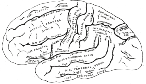
Surface of the cerebral hemispheres in the anterior and middle cranial fossae; the central sulcus separates frontal from parietal lobes, the lateral (Sylvian) fissure separates the temporal lobe below.
- Parts
- Frontal lobe: primary motor cortex (precentral gyrus), premotor and supplementary motor areas, frontal eye fields (voluntary saccades to the opposite side), prefrontal cortex (executive function, judgment, personality), Broca area (inferior frontal gyrus, dominant hemisphere — speech production)
- Parietal lobe: primary somatosensory cortex (postcentral gyrus); dominant inferior parietal lobule (angular and supramarginal gyri — reading, writing, calculation); nondominant parietal lobe (spatial attention — lesion → hemineglect)
- Temporal lobe: primary auditory cortex (Heschl gyrus), Wernicke area (posterior superior temporal gyrus, dominant — language comprehension), medial temporal memory structures (limbic system), Meyer loop of the optic radiation
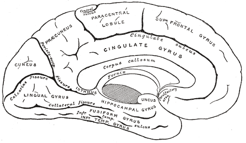
- Occipital lobe: primary visual cortex (V1) around the calcarine sulcus (see optic nerve and visual pathway (CN II))
- Arcuate fasciculus connects Wernicke and Broca areas
- Homunculus: the leg and foot lie on the medial surface (paracentral lobule); the trunk, arm, hand and face lie on the lateral surface, face lowest
- Relations
The left hemisphere is dominant for language in about 95% of right-handers and most left-handers.
- Arterial supply
Anterior cerebral artery (medial frontal and parietal — leg), middle cerebral artery (lateral surface — face, arm, language), posterior cerebral artery (occipital lobe, inferior temporal lobe) — see cerebral arteries (ACA, MCA, PCA).
- Venous drainage
Superficial cerebral veins → superior sagittal and other dural venous sinuses.
Mentioned in
Generated automatically from links in other entries.
Swipe sideways to see the whole diagram →


Clinical correlations
Aphasias PANCE
- Mechanism / lesion
Lesions of the dominant (usually left) hemisphere — most often MCA territory strokes.
- Classic presentation
Broca (expressive): nonfluent, effortful telegraphic speech, comprehension intact, poor repetition, patient aware and frustrated; often with right face/arm weakness (superior MCA division). Wernicke (receptive): fluent but meaningless speech (paraphasias, neologisms), poor comprehension, poor repetition, patient unaware; often no weakness (inferior division). Conduction (arcuate fasciculus): fluent, comprehension intact, repetition poor. Global: all impaired (large MCA stroke).
Parietal lobe syndromes PANCE
- Mechanism / lesion
Stroke, tumor or trauma of the parietal lobe.
- Classic presentation
Nondominant (right): left hemineglect (ignores the left side of space and body), anosognosia, constructional apraxia. Dominant (left) angular gyrus: Gerstmann syndrome — agraphia, acalculia, finger agnosia, left–right confusion. Either side: cortical sensory loss (astereognosis, agraphesthesia).
Frontal lobe syndrome
- Mechanism / lesion
Frontal tumors (e.g. meningioma), trauma, frontotemporal dementia, ACA stroke.
- Classic presentation
Personality change, disinhibition or apathy, poor judgment and planning, primitive reflexes (grasp), urinary incontinence, gaze deviation toward the lesion (frontal eye field) in acute strokes.
Focal seizures
- Mechanism / lesion
Abnormal cortical discharge; symptoms reflect the involved cortex.
- Classic presentation
Motor cortex → clonic jerking that can "march" from hand to face to leg (Jacksonian march), followed by transient weakness (Todd paralysis); occipital → flashing lights; temporal → déjà vu, smells, automatisms.
High-yield PANCE pearls
- Nonfluent speech + intact comprehension → Broca (inferior frontal gyrus).
- Fluent nonsense + poor comprehension → Wernicke (superior temporal gyrus).
- Poor repetition only → conduction aphasia (arcuate fasciculus).
- Left-sided neglect → right parietal lesion.
- Agraphia, acalculia, finger agnosia, L–R confusion → Gerstmann (dominant angular gyrus).
- Leg > arm weakness → ACA; face/arm > leg + aphasia → MCA.
Imaging anatomy
- CT
Noncontrast CT first in acute neurologic deficits (exclude hemorrhage before thrombolysis).
- MRI
MRI with DWI shows acute infarcts within minutes; MRI with contrast for tumors; functional MRI maps language before epilepsy surgery.
- Key views & landmarks
- CT head without contrast (stroke code)
- MRI brain with DWI
- EEG for seizures
Physical exam
- Strength / function testing
Language: fluency, comprehension (one- and multi-step commands), repetition ("no ifs, ands or buts"), naming, reading, writing. Cortical sensation: stereognosis, graphesthesia, two-point discrimination, extinction to double simultaneous stimulation (neglect). Visual fields. Frontal release signs. Cognitive screening (MoCA/MMSE).
Self-quiz
After a stroke, a 67-year-old man speaks in short, halting phrases ("Walk... dog... park"). He follows commands correctly and is clearly frustrated. His right face and arm are weak. Where is the lesion?
Correct answer: B
Nonfluent speech with preserved comprehension and awareness is Broca aphasia, from the left inferior frontal gyrus (superior MCA division), often with right face and arm weakness.
Show answer
B
Nonfluent speech with preserved comprehension and awareness is Broca aphasia, from the left inferior frontal gyrus (superior MCA division), often with right face and arm weakness.
A 72-year-old woman after a stroke eats only from the right side of her plate, ignores people on her left and denies that anything is wrong. Where is the lesion?
Correct answer: B
Hemineglect of the left side with anosognosia (denial of deficit) is typical of a right (nondominant) parietal lesion.
Show answer
B
Hemineglect of the left side with anosognosia (denial of deficit) is typical of a right (nondominant) parietal lesion.
A lesion of the medial surface of the right precentral gyrus would cause weakness mainly of which part of the body?
Correct answer: C
The leg and foot are represented on the medial surface of the motor cortex (ACA territory). Motor output crosses, so a right-sided lesion weakens the left leg.
Show answer
C
The leg and foot are represented on the medial surface of the motor cortex (ACA territory). Motor output crosses, so a right-sided lesion weakens the left leg.
After a stroke, a patient speaks fluently and understands well but cannot repeat a phrase. Which structure is most likely damaged?
Correct answer: D
Conduction aphasia (fluent speech, intact comprehension, poor repetition) results from damage to the arcuate fasciculus connecting Wernicke and Broca areas.
Show answer
D
Conduction aphasia (fluent speech, intact comprehension, poor repetition) results from damage to the arcuate fasciculus connecting Wernicke and Broca areas.
A patient has agraphia, acalculia, finger agnosia and left–right confusion. Where is the lesion?
Correct answer: B
This is Gerstmann syndrome, from a lesion of the dominant (usually left) angular gyrus in the inferior parietal lobule. Nondominant parietal lesions cause hemineglect.
Show answer
B
This is Gerstmann syndrome, from a lesion of the dominant (usually left) angular gyrus in the inferior parietal lobule. Nondominant parietal lesions cause hemineglect.