Glenohumeral joint
Ball-and-socket joint between the humeral head and the glenoid cavity. It is the most mobile joint in the body and the most frequently dislocated major joint.
Core anatomy
- Location
Between the head of the humerus and the glenoid cavity of the scapula, beneath the deltoid and coracoacromial arch.
- Classification
Synovial ball-and-socket (multiaxial) joint.
- Articulating surfaces
Head of the humerus with the glenoid cavity of the scapula. The glenoid is shallow, covering only about a quarter to a third of the head, and is deepened by the fibrocartilaginous glenoid labrum.
- Capsule
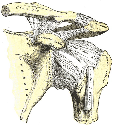
Loose fibrous capsule from the margin of the glenoid to the anatomical neck of the humerus (extending further down the shaft inferiorly). It is weakest inferiorly, where there is no rotator cuff support. The long head of biceps brachii passes through the joint (intracapsular, enclosed in a synovial sheath) to reach the intertubercular groove.
- Ligaments
- Glenohumeral ligaments (superior, middle, inferior): thickenings of the anterior capsule; the inferior glenohumeral ligament is the key anterior stabilizer in abduction–external rotation
- Coracohumeral ligament: coracoid base → greater tubercle; strengthens the superior capsule
- Transverse humeral ligament: bridges the intertubercular groove, holding the biceps tendon
- Coracoacromial ligament: with the acromion and coracoid forms the coracoacromial arch, which prevents superior displacement of the head
- Movements
- Flexion–extension, abduction–adduction, medial–lateral rotation, circumduction
- Full elevation combines glenohumeral and scapulothoracic motion (scapulohumeral rhythm, about 2:1 glenohumeral to scapular) verify
- Stability
Mobility comes at the expense of stability. Dynamic stability comes mainly from the rotator cuff (supraspinatus, infraspinatus, teres minor, subscapularis) and the long head of biceps. Static stability comes from the labrum, capsule, glenohumeral ligaments and coracoacromial arch.
- Innervation
Suprascapular, axillary nerve and lateral pectoral nerves
- Blood supply
Anterior and posterior circumflex humeral arteries and branches of the suprascapular artery (from the axillary artery and subclavian systems)
- Relations
The subacromial–subdeltoid bursa lies superiorly. The subscapular bursa (anterior) often communicates with the joint. The axillary nerve and posterior circumflex humeral artery pass directly below the capsule.
Mentioned in
Generated automatically from links in other entries.
Swipe sideways to see the whole diagram →

Clinical correlations
Anterior dislocation PANCE
- Mechanism / lesion
By far the most common direction. Caused by abduction + external rotation (with extension), e.g. blocking a ball overhead or a fall. The head moves anteroinferiorly to a subcoracoid position.
- Classic presentation
Squared-off shoulder (loss of the deltoid contour, prominent acromion). The arm is held slightly abducted and externally rotated, and the patient resists moving it. Check the axillary nerve (regimental badge sensation). Associated injuries include the Bankart lesion (anteroinferior labral tear ± glenoid rim fracture) and the Hill–Sachs lesion (impaction of the posterolateral humeral head). Recurrence is common in young patients.
Posterior dislocation PANCE
- Mechanism / lesion
Classically seizures or electric shock (strong internal rotators overpower external rotators), or a direct anterior blow.
- Classic presentation
Arm held adducted and internally rotated; the patient can't externally rotate. Posterior fullness and anterior flattening. Easily missed on a single AP view. Reverse Hill–Sachs lesion.
Adhesive capsulitis (frozen shoulder) PANCE
- Mechanism / lesion
Inflammation and fibrosis of the capsule. Associated with diabetes mellitus, thyroid disease and immobilization. Typically ages 40–60, more often women.
- Classic presentation
Painful, then stiff, then "thawing" phases. Loss of both active and passive ROM, most markedly external rotation. Radiographs are normal.
Glenohumeral osteoarthritis
- Mechanism / lesion
Degenerative loss of articular cartilage, often in older adults or after prior instability or trauma.
- Classic presentation
Deep shoulder pain, stiffness and crepitus, with loss of external rotation. Radiographs show joint space narrowing, an inferior humeral osteophyte and subchondral sclerosis.
High-yield PANCE pearls
- Anterior dislocation (≈95%): abduction + external rotation; squared-off shoulder; axillary nerve; Bankart + Hill–Sachs.
- Posterior dislocation: seizure / electrocution; arm adducted and internally rotated; lightbulb sign on AP.
- Adhesive capsulitis: diabetes; loss of active and passive ROM (especially external rotation).
- Rotator cuff = the main dynamic stabilizer; inferior capsule = the weakest region.
Imaging anatomy
- X-ray
Trauma series: AP (or Grashey), scapular Y and axillary view. On the Y view, the head should overlie the center of the Y. Anterior dislocation: head subcoracoid. Posterior dislocation: the AP view may show a fixed internally rotated head ("lightbulb sign") and widening of the space between the anterior glenoid rim and the head ("rim sign"); the axillary view is confirmatory. Hill–Sachs lesions are best seen on an AP view with internal rotation.
- CT
Quantifies glenoid bone loss (bony Bankart) and Hill–Sachs size in recurrent instability; complex fractures.
- MRI
MR arthrography (intra-articular contrast) is the reference test for labral tears (Bankart, SLAP) and capsular injury. Non-contrast MRI evaluates the rotator cuff, bone bruising and osteonecrosis. In adhesive capsulitis, capsular thickening in the axillary recess and rotator interval is described.
- Ultrasound
Dynamic evaluation of the rotator cuff, biceps tendon, effusion and subacromial bursa. Guides joint injections.
- Key views & landmarks
- AP/Grashey, scapular Y, axillary lateral
- MR arthrogram for labral pathology
Physical exam
- Surface landmarks
Acromion (point of the shoulder), greater tubercle (lateral, beneath deltoid), coracoid process (deltopectoral triangle), bicipital groove (anterior, between the tubercles).
- Palpation
Palpate the AC joint, acromion, greater tubercle, bicipital groove and coracoid for tenderness. Inspect the contour from front and back.
- Strength / function testing
Active and passive ROM in all planes (comparing the two isolates capsular vs cuff pathology). Cuff strength tests. Neurovascular exam (axillary nerve) before and after any reduction.
Apprehension test
- Technique
Patient supine; the shoulder is abducted to 90° and the examiner gradually externally rotates it.
- Positive test
The patient becomes apprehensive or resists, fearing dislocation (pain alone is less specific).
- Significance
Anterior instability.
Relocation test
- Technique
Performed after a positive apprehension test. The examiner applies a posteriorly directed force to the humeral head.
- Positive test
Apprehension is relieved and further external rotation is tolerated.
- Significance
Confirms anterior instability.
Sulcus sign
- Technique
With the arm relaxed at the side, the examiner pulls the arm inferiorly.
- Positive test
A visible depression (sulcus) appears between the acromion and the humeral head.
- Significance
Inferior or multidirectional instability.
Self-quiz
A 24-year-old man is brought in after a generalized tonic-clonic seizure with right shoulder pain. He holds the arm adducted and internally rotated and can't externally rotate it. The AP radiograph shows a symmetric, rounded humeral head. What is the most likely diagnosis?
Correct answer: B
Seizures cause posterior dislocation. The arm is locked in internal rotation, and the AP view shows the "lightbulb sign". Always obtain an axillary or scapular Y view, because posterior dislocations are frequently missed on AP alone.
Show answer
B
Seizures cause posterior dislocation. The arm is locked in internal rotation, and the AP view shows the "lightbulb sign". Always obtain an axillary or scapular Y view, because posterior dislocations are frequently missed on AP alone.
A 54-year-old woman with type 2 diabetes has 5 months of progressive shoulder pain and stiffness. Both active and passive external rotation are markedly reduced. Radiographs are normal. What is the most likely diagnosis?
Correct answer: C
Adhesive capsulitis restricts both active and passive motion, especially external rotation, and is associated with diabetes. A cuff tear limits active motion while passive motion is preserved. OA would show radiographic changes.
Show answer
C
Adhesive capsulitis restricts both active and passive motion, especially external rotation, and is associated with diabetes. A cuff tear limits active motion while passive motion is preserved. OA would show radiographic changes.
Which lesion is an impaction fracture of the posterolateral humeral head after anterior dislocation?
Correct answer: B
The Hill–Sachs lesion forms when the posterolateral head impacts the anterior glenoid rim. The Bankart lesion is the anteroinferior labral (± glenoid rim) injury. A reverse Hill–Sachs (anteromedial head) follows posterior dislocation.
Show answer
B
The Hill–Sachs lesion forms when the posterolateral head impacts the anterior glenoid rim. The Bankart lesion is the anteroinferior labral (± glenoid rim) injury. A reverse Hill–Sachs (anteromedial head) follows posterior dislocation.
What is the main dynamic stabilizer of the glenohumeral joint?
Correct answer: C
The rotator cuff muscles compress the humeral head into the glenoid throughout motion. The labrum, ligaments and arch are static stabilizers.
Show answer
C
The rotator cuff muscles compress the humeral head into the glenoid throughout motion. The labrum, ligaments and arch are static stabilizers.
After an anterior shoulder dislocation, MRI shows a tear of the anteroinferior glenoid labrum. What is this lesion called?
Correct answer: C
A Bankart lesion is an anteroinferior labral tear (± glenoid rim fracture, a "bony Bankart") after anterior dislocation. The Hill–Sachs lesion is the matching impaction of the posterolateral humeral head. A reverse Hill–Sachs follows posterior dislocation; a SLAP lesion is a superior labral tear.
Show answer
C
A Bankart lesion is an anteroinferior labral tear (± glenoid rim fracture, a "bony Bankart") after anterior dislocation. The Hill–Sachs lesion is the matching impaction of the posterolateral humeral head. A reverse Hill–Sachs follows posterior dislocation; a SLAP lesion is a superior labral tear.